Back


Poster Session A - Sunday Afternoon
Category: General Endoscopy
A0283 - Diffuse Mesenteric Lymphadenopathy and Duodenal Polyposis Secondary to Mantle Cell Lymphoma: A Rare Case Report
Sunday, October 23, 2022
5:00 PM – 7:00 PM ET
Location: Crown Ballroom

Has Audio

Rami Musallam, MD
St. Vincent Charity Medical Center
Cleveland, OH
Presenting Author(s)
Rami Musallam, MD1, Serge Matar, MD2, Noor Aldeen Abdel Jalil, 3, Ala Abdel Jalil, MD4
1St. Vincent Charity Medical Center, Cleveland, OH; 2St. Joseph University, Cleveland, OH; 3University of Jordan, Cleveland, OH; 4Cleveland Clinic Fairview Hospital, Cleveland, OH
Introduction: Mantle cell lymphoma (MCL) is a rare type of B-cell non-Hodgkin lymphoma characterized by atypical small lymphoid cells within the mantle zone of germinal center follicles. Immunohistochemistry shows cyclin D1 overexpression associated with CD5+ and CD20+ expression and a t(11;34)(q13;q32) translocation. Despite being classified as low-grade, it is an aggressive lymphoma because it is usually discovered at a late stage with splenomegaly, lymphadenopathy, and blood and bone marrow involvement. Gastrointestinal involvement is common as an extranodal site of the MCL; however, primary GI lymphoma is very rare (about 1-4% of all GI malignancies).
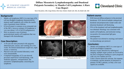
Case Description/Methods: A 58-year-old male presented with epigastric abdominal pain, nausea, and vomiting for a few weeks. He denied unintentional weight loss, night sweats, or anorexia. The physical exam and essential laboratory work were unremarkable. Computed tomography (CT) scan showed proximal small bowel wall thickening, with moderate epigastric, retroperitoneal, and mesenteric lymphadenopathy concerning lymphoma. Esophagogastroduodenoscopy (EGD) showed diffuse polyposis in the proximal duodenum (Figure1 A,B). Endoscopic ultrasound (EUS) showed multiple enlarged peri duodenal and peripancreatic lymph nodes and abnormal duodenal wall thickening. A fine needle biopsy of the duodenal wall and lymphadenopathy was obtained using a 22-gauge needle (Figure1 C,D). Histology was consistent with mantle cell lymphoma, and molecular testing was positive for monoclonal IgH gene rearrangement. The patient was referred to oncology and started on chemotherapy per protocol.
Discussion: Mantle cell lymphoma (MCL) is a rare type of B-cell non-Hodgkin lymphoma. Because of poor detection by radiological imaging, multiple organ systems are involved by the time of diagnosis leading to a poor prognosis with very low median survival. Only a few case reports are available in the literature about primary gastrointestinal mantle cell lymphoma; thus, little is known about the outcome, the response to treatment, and the duration of remission in primary GI MCL patients.

Disclosures:
Rami Musallam, MD1, Serge Matar, MD2, Noor Aldeen Abdel Jalil, 3, Ala Abdel Jalil, MD4. A0283 - Diffuse Mesenteric Lymphadenopathy and Duodenal Polyposis Secondary to Mantle Cell Lymphoma: A Rare Case Report, ACG 2022 Annual Scientific Meeting Abstracts. Charlotte, NC: American College of Gastroenterology.
1St. Vincent Charity Medical Center, Cleveland, OH; 2St. Joseph University, Cleveland, OH; 3University of Jordan, Cleveland, OH; 4Cleveland Clinic Fairview Hospital, Cleveland, OH
Introduction: Mantle cell lymphoma (MCL) is a rare type of B-cell non-Hodgkin lymphoma characterized by atypical small lymphoid cells within the mantle zone of germinal center follicles. Immunohistochemistry shows cyclin D1 overexpression associated with CD5+ and CD20+ expression and a t(11;34)(q13;q32) translocation. Despite being classified as low-grade, it is an aggressive lymphoma because it is usually discovered at a late stage with splenomegaly, lymphadenopathy, and blood and bone marrow involvement. Gastrointestinal involvement is common as an extranodal site of the MCL; however, primary GI lymphoma is very rare (about 1-4% of all GI malignancies).
Case Description/Methods: A 58-year-old male presented with epigastric abdominal pain, nausea, and vomiting for a few weeks. He denied unintentional weight loss, night sweats, or anorexia. The physical exam and essential laboratory work were unremarkable. Computed tomography (CT) scan showed proximal small bowel wall thickening, with moderate epigastric, retroperitoneal, and mesenteric lymphadenopathy concerning lymphoma. Esophagogastroduodenoscopy (EGD) showed diffuse polyposis in the proximal duodenum (Figure1 A,B). Endoscopic ultrasound (EUS) showed multiple enlarged peri duodenal and peripancreatic lymph nodes and abnormal duodenal wall thickening. A fine needle biopsy of the duodenal wall and lymphadenopathy was obtained using a 22-gauge needle (Figure1 C,D). Histology was consistent with mantle cell lymphoma, and molecular testing was positive for monoclonal IgH gene rearrangement. The patient was referred to oncology and started on chemotherapy per protocol.
Discussion: Mantle cell lymphoma (MCL) is a rare type of B-cell non-Hodgkin lymphoma. Because of poor detection by radiological imaging, multiple organ systems are involved by the time of diagnosis leading to a poor prognosis with very low median survival. Only a few case reports are available in the literature about primary gastrointestinal mantle cell lymphoma; thus, little is known about the outcome, the response to treatment, and the duration of remission in primary GI MCL patients.

Figure: Figure 1: (A) EGD image shows diffuse polyposis in the proximal duodenum, (B) EGD image with narrow-band imaging shows diffuse duodenal polyposis, (C) Endoscopic ultrasound shows peripancreatic lymphadenopathy, (D) Endoscopic ultrasound with fine-needle biopsy of the thickened duodenal wall.
Disclosures:
Rami Musallam indicated no relevant financial relationships.
Serge Matar indicated no relevant financial relationships.
Noor Aldeen Abdel Jalil indicated no relevant financial relationships.
Ala Abdel Jalil indicated no relevant financial relationships.
Rami Musallam, MD1, Serge Matar, MD2, Noor Aldeen Abdel Jalil, 3, Ala Abdel Jalil, MD4. A0283 - Diffuse Mesenteric Lymphadenopathy and Duodenal Polyposis Secondary to Mantle Cell Lymphoma: A Rare Case Report, ACG 2022 Annual Scientific Meeting Abstracts. Charlotte, NC: American College of Gastroenterology.
