Back


Poster Session E - Tuesday Afternoon
Category: Small Intestine
E0665 - Capsule Endoscopy as a Diagnostic Tool for Metastatic Melanoma of the Small Bowel
Tuesday, October 25, 2022
3:00 PM – 5:00 PM ET
Location: Crown Ballroom

Has Audio

Navjit Singh, MD, MSc
University of Illinois College of Medicine at Peoria
Peoria, IL
Presenting Author(s)
Navjit Singh, MD, MSc, Mohammad Gareeb, MD, MBBS, Yeshaswini Panathur Sreenivasa Reddy, MD, Imran Balouch, MD
University of Illinois College of Medicine at Peoria, Peoria, IL
Introduction: The detection of gastrointestinal malignancies in the small bowel remains to be a challenge for both clinicians and radiologists due to nonspecific clinical findings and inconclusive diagnostic testing. Endoluminal studies are beneficial but have their limitations on particular areas of bowel. Capsule endoscopy is a noninvasive imaging study with a high yield in diagnosing these cases.
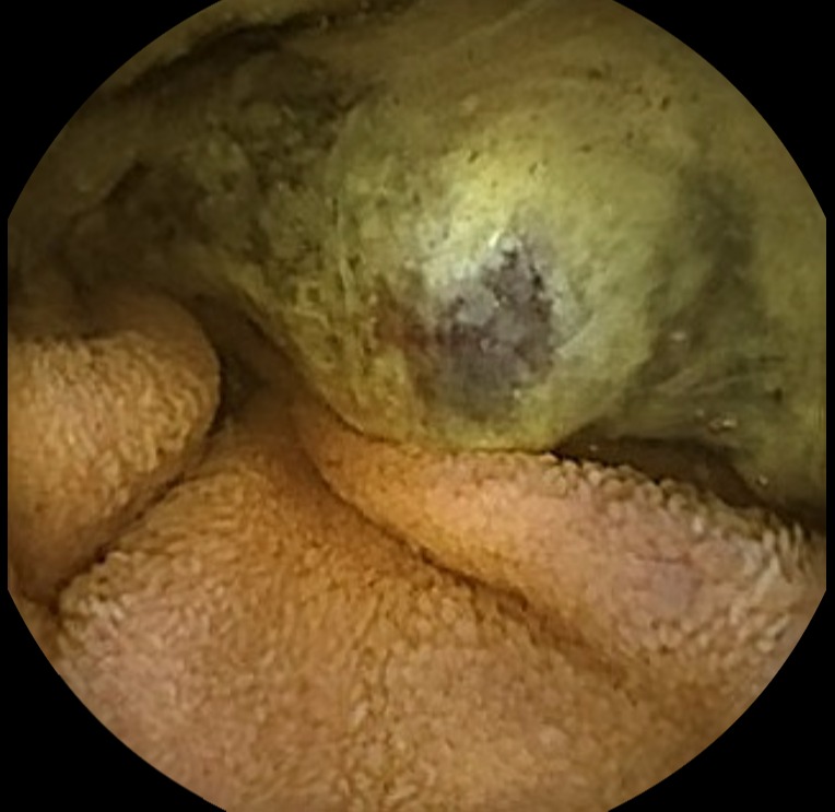
Case Description/Methods: A 53 year-old male with a history of melanoma of the back was treated with surgical excision and immunotherapy presented with a 2 month history of progressive exertional dyspnea and fatigue. Denied any complaints of overt bleeding, abdominal pain, or weight loss. No family history of gastrointestinal malignancies. Noted to have iron deficiency anemia with a hemoglobin of 6.7 g/dL. He was admitted and received blood transfusion with improvement of his symptoms and wanted to pursue outpatient endoscopies. However continued to have ongoing symptoms following discharge. He underwent an upper endoscopy which was unremarkable and a colonoscopy that showed a small transverse colon polyp and small internal hemorrhoids which did not explain his anemia. Therefore he underwent a small bowel capsule endoscopy that revealed a partially obstructing ulcerated mass with active oozing in the proximal small bowel, seen in fig. 1. He underwent an urgent upper endoscopy with push enteroscopy that demonstrated a large fungating multilobulated jejunal mass that was biopsied. Pathology confirmed the mass to be a MART-1, S100, and HMB45 positive melanoma, suspected to be a metastatic lesion given his history. A CT scan of his abdomen and pelvis was done for staging purposes that showed new metastatic tumoral implants in the mesentery adjacent to the small bowel loops.
Discussion: Malignant melanomas in the gastrointestinal tract are rare and could be either a true primary or a metastatic lesion. They are difficult to diagnose as only 1-5% are detected in early stages of imaging. Routine endoscopic modalities have minimal benefit in diagnosing small bowel lesions. These malignancies are aggressive and are diagnosed in late stages with conventional studies and have a poor prognosis. Therefore capsule endoscopy is a simple and noninvasive test that can help provide crucial information in establishing diagnosis.

Disclosures:
Navjit Singh, MD, MSc, Mohammad Gareeb, MD, MBBS, Yeshaswini Panathur Sreenivasa Reddy, MD, Imran Balouch, MD. E0665 - Capsule Endoscopy as a Diagnostic Tool for Metastatic Melanoma of the Small Bowel, ACG 2022 Annual Scientific Meeting Abstracts. Charlotte, NC: American College of Gastroenterology.
University of Illinois College of Medicine at Peoria, Peoria, IL
Introduction: The detection of gastrointestinal malignancies in the small bowel remains to be a challenge for both clinicians and radiologists due to nonspecific clinical findings and inconclusive diagnostic testing. Endoluminal studies are beneficial but have their limitations on particular areas of bowel. Capsule endoscopy is a noninvasive imaging study with a high yield in diagnosing these cases.
Case Description/Methods: A 53 year-old male with a history of melanoma of the back was treated with surgical excision and immunotherapy presented with a 2 month history of progressive exertional dyspnea and fatigue. Denied any complaints of overt bleeding, abdominal pain, or weight loss. No family history of gastrointestinal malignancies. Noted to have iron deficiency anemia with a hemoglobin of 6.7 g/dL. He was admitted and received blood transfusion with improvement of his symptoms and wanted to pursue outpatient endoscopies. However continued to have ongoing symptoms following discharge. He underwent an upper endoscopy which was unremarkable and a colonoscopy that showed a small transverse colon polyp and small internal hemorrhoids which did not explain his anemia. Therefore he underwent a small bowel capsule endoscopy that revealed a partially obstructing ulcerated mass with active oozing in the proximal small bowel, seen in fig. 1. He underwent an urgent upper endoscopy with push enteroscopy that demonstrated a large fungating multilobulated jejunal mass that was biopsied. Pathology confirmed the mass to be a MART-1, S100, and HMB45 positive melanoma, suspected to be a metastatic lesion given his history. A CT scan of his abdomen and pelvis was done for staging purposes that showed new metastatic tumoral implants in the mesentery adjacent to the small bowel loops.
Discussion: Malignant melanomas in the gastrointestinal tract are rare and could be either a true primary or a metastatic lesion. They are difficult to diagnose as only 1-5% are detected in early stages of imaging. Routine endoscopic modalities have minimal benefit in diagnosing small bowel lesions. These malignancies are aggressive and are diagnosed in late stages with conventional studies and have a poor prognosis. Therefore capsule endoscopy is a simple and noninvasive test that can help provide crucial information in establishing diagnosis.

Figure: Figure 1: Video Capsule Endoscopy demonstrating an ulcerated mass visualized in the jejunum with partial obstruction.
Disclosures:
Navjit Singh indicated no relevant financial relationships.
Mohammad Gareeb indicated no relevant financial relationships.
Yeshaswini Panathur Sreenivasa Reddy indicated no relevant financial relationships.
Imran Balouch indicated no relevant financial relationships.
Navjit Singh, MD, MSc, Mohammad Gareeb, MD, MBBS, Yeshaswini Panathur Sreenivasa Reddy, MD, Imran Balouch, MD. E0665 - Capsule Endoscopy as a Diagnostic Tool for Metastatic Melanoma of the Small Bowel, ACG 2022 Annual Scientific Meeting Abstracts. Charlotte, NC: American College of Gastroenterology.
