Back


Poster Session E - Tuesday Afternoon
Category: Stomach
E0712 - Gastric Neuroendocrine Tumor of Indeterminate Type
Tuesday, October 25, 2022
3:00 PM – 5:00 PM ET
Location: Crown Ballroom

Has Audio

Michael Tran, MD
University of South Alabama
Mobile, AL
Presenting Author(s)
Michael Tran, MD, Hajira Zafar Malik, MD, Chad Spencer, MD, Phillip Henderson, DO
University of South Alabama, Mobile, AL
Introduction: Gastric neuroendocrine tumors (GNETs) are rare tumors making up less than 2% of gastric polyps. Type I GNETs are most common (70-80%) and are associated with atrophic gastritis, pernicious anemia, iron deficiency, elevated gastrin, and high gastric pH. These are typically small, low-grade, and may be multifocal. Type II tumors are also associated with elevated gastrin levels but a low pH, as seen in Zollinger-Ellison syndrome. Type III tumors are larger and more aggressive and occur sporadically in the setting of normal gastric mucosa, normal gastrin levels, and normal pH. Here we discuss an unusual case of GNET which does not fit into either category.
Case Description/Methods: A 70-year-old white male with history of gastroesophageal reflux disease (GERD) on chronic proton pump inhibitor (PPI) presented for esophagogastroduodenoscopy (EGD) for worsening GERD symptoms. EGD revealed 2 polyps in the body – each was 4-5 mm and was removed with hot snare. A few smaller polyps were removed with biopsy forceps. The gastric mucosa was grossly normal. Sydney protocol biopsies were obtained. The 2 larger polyps returned as well-differentiated GNETs, grade 1 (Ki-67 index 2.5%). The smaller polyps returned as areas of intestinal metaplasia (IM), but Sydney protocol biopsies did not reveal H. pylori, atrophic gastritis, or IM. EGD was repeated off PPI for 1 week and fasting labs were obtained. Gastric pH was 1, serum gastrin was 78 pg/mL (normal), vitamin B12 and ferritin were normal, and intrinsic factor and parietal cell antibodies were negative.
Discussion: This case highlights a scenario in which a GNET is difficult to categorize based on classic criteria. As type III lesions are very aggressive with high risk of metastasis, distinguishing between type I and type III lesions is paramount. While grossly and histologically resembling a type I GNET (multifocal, small, low-grade), the GNETs in this case were present in the absence of atrophic gastritis, and the patient had a normal gastrin and low gastric pH off PPI. The lack of atrophic gastritis and normal gastrin levels are more suggestive of a type III tumor, but one would expect a single, large, high-grade GNET in that case.
As PPI use has increased in the past 30 years, so has incidence of GNETs; it is possible that a chronic, PPI-induced “hyper-gastrin” state was responsible for this patient’s GNETs and that gastrin normalized once PPI was discontinued. More studies are needed to further categorize GNETs with indeterminate features.

Disclosures:
Michael Tran, MD, Hajira Zafar Malik, MD, Chad Spencer, MD, Phillip Henderson, DO. E0712 - Gastric Neuroendocrine Tumor of Indeterminate Type, ACG 2022 Annual Scientific Meeting Abstracts. Charlotte, NC: American College of Gastroenterology.
University of South Alabama, Mobile, AL
Introduction: Gastric neuroendocrine tumors (GNETs) are rare tumors making up less than 2% of gastric polyps. Type I GNETs are most common (70-80%) and are associated with atrophic gastritis, pernicious anemia, iron deficiency, elevated gastrin, and high gastric pH. These are typically small, low-grade, and may be multifocal. Type II tumors are also associated with elevated gastrin levels but a low pH, as seen in Zollinger-Ellison syndrome. Type III tumors are larger and more aggressive and occur sporadically in the setting of normal gastric mucosa, normal gastrin levels, and normal pH. Here we discuss an unusual case of GNET which does not fit into either category.
Case Description/Methods: A 70-year-old white male with history of gastroesophageal reflux disease (GERD) on chronic proton pump inhibitor (PPI) presented for esophagogastroduodenoscopy (EGD) for worsening GERD symptoms. EGD revealed 2 polyps in the body – each was 4-5 mm and was removed with hot snare. A few smaller polyps were removed with biopsy forceps. The gastric mucosa was grossly normal. Sydney protocol biopsies were obtained. The 2 larger polyps returned as well-differentiated GNETs, grade 1 (Ki-67 index 2.5%). The smaller polyps returned as areas of intestinal metaplasia (IM), but Sydney protocol biopsies did not reveal H. pylori, atrophic gastritis, or IM. EGD was repeated off PPI for 1 week and fasting labs were obtained. Gastric pH was 1, serum gastrin was 78 pg/mL (normal), vitamin B12 and ferritin were normal, and intrinsic factor and parietal cell antibodies were negative.
Discussion: This case highlights a scenario in which a GNET is difficult to categorize based on classic criteria. As type III lesions are very aggressive with high risk of metastasis, distinguishing between type I and type III lesions is paramount. While grossly and histologically resembling a type I GNET (multifocal, small, low-grade), the GNETs in this case were present in the absence of atrophic gastritis, and the patient had a normal gastrin and low gastric pH off PPI. The lack of atrophic gastritis and normal gastrin levels are more suggestive of a type III tumor, but one would expect a single, large, high-grade GNET in that case.
As PPI use has increased in the past 30 years, so has incidence of GNETs; it is possible that a chronic, PPI-induced “hyper-gastrin” state was responsible for this patient’s GNETs and that gastrin normalized once PPI was discontinued. More studies are needed to further categorize GNETs with indeterminate features.

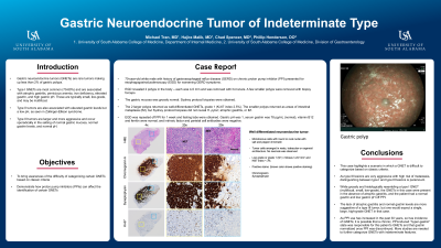
Figure: Gastric Polyp
Disclosures:
Michael Tran indicated no relevant financial relationships.
Hajira Zafar Malik indicated no relevant financial relationships.
Chad Spencer indicated no relevant financial relationships.
Phillip Henderson indicated no relevant financial relationships.
Michael Tran, MD, Hajira Zafar Malik, MD, Chad Spencer, MD, Phillip Henderson, DO. E0712 - Gastric Neuroendocrine Tumor of Indeterminate Type, ACG 2022 Annual Scientific Meeting Abstracts. Charlotte, NC: American College of Gastroenterology.
