Back


Poster Session E - Tuesday Afternoon
Category: Stomach
E0716 - Where Is the Bumper? A Case of Percutaneous Endoscopic Gastrostomy (PEG) Tube Malfunction
Tuesday, October 25, 2022
3:00 PM – 5:00 PM ET
Location: Crown Ballroom

Has Audio
- TB
Tim Brotherton, MD
Saint Louis University
St. Louis, Missouri
Presenting Author(s)
Tim Brotherton, MD1, Laith Numan, MD1, Gregory Sayuk, MD2, Michael Presti, MD3, Jill E. Elwing, MD2
1Saint Louis University, St. Louis, MO; 2St. Louis VA, Washington University, St. Louis, MO; 3St. Louis VA, Saint Louis University, St. Louis, MO
Introduction: Percutaneous endoscopic gastrostomy (PEG) tubes are used for enteral access in patients requiring nutritional support. PEG tube placement and removal generally are regarded as safe procedures. However, complications can be experienced in 16-70%, ranging from benign pneumoperitoneum to gastric outlet obstruction, buried bumper syndrome, and bowel perforation. Our case describes a retained internal bumper during PEG tube removal that undergoes spontaneous passage.
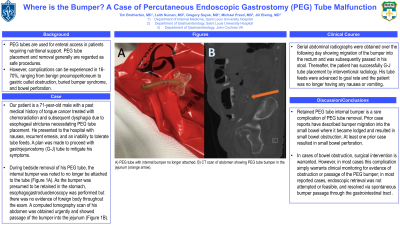
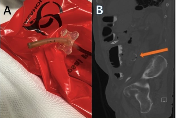
Case Description/Methods: Our patient is a 71-year-old male with a past medical history of tongue cancer treated with chemoradiation and subsequent dysphagia due to esophageal strictures necessitating PEG tube placement. He presented to the hospital with nausea, recurrent emesis, and an inability to tolerate tube feeds. His PEG tube was placed to gravity with one liter of output and relief of symptoms. A plan was made to proceed with gastrojejunostomy (G-J) tube to mitigate his symptoms. During bedside removal of his PEG tube, the internal bumper was noted to no longer be attached to the tube (Figure 1A). A Foley catheter was placed in the tract to maintain its patency. As the bumper was presumed to be retained in the stomach, esophagogastroduodenoscopy was performed but there was no evidence of foreign body throughout the exam. A computed tomography scan of his abdomen was obtained urgently and showed passage of the bumper into the jejunum (Figure 1B).
Serial abdominal radiographs were obtained over the following day showing migration of the bumper into the rectum and was subsequently passed in his stool. Thereafter, the patient has successfully G-J tube placement by interventional radiology. His tube feeds were advanced to goal rate and the patient was no longer having any nausea or vomiting.
Discussion: Retained PEG tube internal bumper is a rare complication of PEG tube removal. Prior case reports have described bumper migration into the small bowel where it became lodged and resulted in small bowel obstruction. At least one prior case resulted in small bowel perforation. In cases of bowel obstruction, surgical intervention is warranted. However, in most cases this complication simply warrants clinical monitoring for evidence of obstruction or passage of the PEG bumper; in most reported cases, endoscopic retrieval was not attempted or feasible, and resolved via spontaneous bumper passage through the gastrointestinal tract .

Disclosures:
Tim Brotherton, MD1, Laith Numan, MD1, Gregory Sayuk, MD2, Michael Presti, MD3, Jill E. Elwing, MD2. E0716 - Where Is the Bumper? A Case of Percutaneous Endoscopic Gastrostomy (PEG) Tube Malfunction, ACG 2022 Annual Scientific Meeting Abstracts. Charlotte, NC: American College of Gastroenterology.
1Saint Louis University, St. Louis, MO; 2St. Louis VA, Washington University, St. Louis, MO; 3St. Louis VA, Saint Louis University, St. Louis, MO
Introduction: Percutaneous endoscopic gastrostomy (PEG) tubes are used for enteral access in patients requiring nutritional support. PEG tube placement and removal generally are regarded as safe procedures. However, complications can be experienced in 16-70%, ranging from benign pneumoperitoneum to gastric outlet obstruction, buried bumper syndrome, and bowel perforation. Our case describes a retained internal bumper during PEG tube removal that undergoes spontaneous passage.
Case Description/Methods: Our patient is a 71-year-old male with a past medical history of tongue cancer treated with chemoradiation and subsequent dysphagia due to esophageal strictures necessitating PEG tube placement. He presented to the hospital with nausea, recurrent emesis, and an inability to tolerate tube feeds. His PEG tube was placed to gravity with one liter of output and relief of symptoms. A plan was made to proceed with gastrojejunostomy (G-J) tube to mitigate his symptoms. During bedside removal of his PEG tube, the internal bumper was noted to no longer be attached to the tube (Figure 1A). A Foley catheter was placed in the tract to maintain its patency. As the bumper was presumed to be retained in the stomach, esophagogastroduodenoscopy was performed but there was no evidence of foreign body throughout the exam. A computed tomography scan of his abdomen was obtained urgently and showed passage of the bumper into the jejunum (Figure 1B).
Serial abdominal radiographs were obtained over the following day showing migration of the bumper into the rectum and was subsequently passed in his stool. Thereafter, the patient has successfully G-J tube placement by interventional radiology. His tube feeds were advanced to goal rate and the patient was no longer having any nausea or vomiting.
Discussion: Retained PEG tube internal bumper is a rare complication of PEG tube removal. Prior case reports have described bumper migration into the small bowel where it became lodged and resulted in small bowel obstruction. At least one prior case resulted in small bowel perforation. In cases of bowel obstruction, surgical intervention is warranted. However, in most cases this complication simply warrants clinical monitoring for evidence of obstruction or passage of the PEG bumper; in most reported cases, endoscopic retrieval was not attempted or feasible, and resolved via spontaneous bumper passage through the gastrointestinal tract .

Figure: A) PEG tube with internal bumper no longer attached.
B) CT scan of abdomen showing PEG tube bumper in the jejunum (orange arrow).
B) CT scan of abdomen showing PEG tube bumper in the jejunum (orange arrow).
Disclosures:
Tim Brotherton indicated no relevant financial relationships.
Laith Numan indicated no relevant financial relationships.
Gregory Sayuk indicated no relevant financial relationships.
Michael Presti indicated no relevant financial relationships.
Jill Elwing indicated no relevant financial relationships.
Tim Brotherton, MD1, Laith Numan, MD1, Gregory Sayuk, MD2, Michael Presti, MD3, Jill E. Elwing, MD2. E0716 - Where Is the Bumper? A Case of Percutaneous Endoscopic Gastrostomy (PEG) Tube Malfunction, ACG 2022 Annual Scientific Meeting Abstracts. Charlotte, NC: American College of Gastroenterology.
