Back


Poster Session E - Tuesday Afternoon
Category: Stomach
E0719 - An Unusual Presentation of a Large Gastrointestinal Stromal Tumor (GIST)
Tuesday, October 25, 2022
3:00 PM – 5:00 PM ET
Location: Crown Ballroom

Has Audio

Jamil M. Shah, MD
The Brooklyn Hospital Center
Brooklyn, NY
Presenting Author(s)
Jamil M. Shah, MD, Praneeth Bandaru, MD, Vijay Gayam, MD, Vikash Kumar, MD, Philip Xiao, MD, Madhavi Reddy, MD, Denzil Etienne, MD
The Brooklyn Hospital Center, Brooklyn, NY
Introduction: Gastrointestinal stromal tumors (GISTs), although the most common mesenchymal neoplasms of the digestive tract, are relatively rare primary GI cancers. They originate from the interstitial cells of Cajal, which are cells of the intestinal autonomic nervous system. They function as electrical pacemakers, controlling motility as well as regulating peristalsis. Individuals with GISTs can present with overt or occult GI bleeding, but, more frequently, they present with nonspecific symptoms, such as vague abdominal pain or discomfort, early satiety, or bloating. Other individuals may be asymptomatic, and the GISTs are detected incidentally during an endoscopic study (where they usually appear as subepithelial masses) or on cross-sectional imaging studies performed for a different reason. Here, we present an interesting case of an elderly woman discovered to have a large GIST.
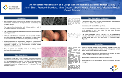
Case Description/Methods: A 69 y/o F, with PMH of b/l unprovoked pulmonary embolism (submassive with evidence of right heart strain) in 8/2017 on anticoagulation (Warfarin), HTN, and DM, underwent EGD and EUS a few years prior that showed a 4 cm umbilicated, firm, round mass along the posterior wall/greater curve of the stomach. The pathology from FNB at the time was suggestive of gastrointestinal stromal tumor (GIST) (+ C-kit, CD117). She subsequently failed to follow up with GI. Recent CT A/P then showed a small hiatal hernia, and a filling defect in the stomach, 3.2x3.1cm, arising from the lesser curvature immediately distal to the GEJ. The patient underwent EGD on 1/12/2022 for intraoperative evaluation of GIST tumor (Fig 1A), followed by robotic assisted surgical wedge resection of the gastric tumor by the surgical team (Fig 1B-E).
Discussion: GISTs are identified primarily by the expression of the KIT protein and often carry activating mutations in either the KIT or the platelet-derived growth factor receptor alpha (PDGFRA) genes.1,3 These neoplasms are frequently discovered in the stomach (40 to 60 percent) and small intestine (30 to 35 percent).4 However, they can arise in any part of the digestive tract. Resectable GISTs can be completely or almost completely removed by surgery.

Disclosures:
Jamil M. Shah, MD, Praneeth Bandaru, MD, Vijay Gayam, MD, Vikash Kumar, MD, Philip Xiao, MD, Madhavi Reddy, MD, Denzil Etienne, MD. E0719 - An Unusual Presentation of a Large Gastrointestinal Stromal Tumor (GIST), ACG 2022 Annual Scientific Meeting Abstracts. Charlotte, NC: American College of Gastroenterology.
The Brooklyn Hospital Center, Brooklyn, NY
Introduction: Gastrointestinal stromal tumors (GISTs), although the most common mesenchymal neoplasms of the digestive tract, are relatively rare primary GI cancers. They originate from the interstitial cells of Cajal, which are cells of the intestinal autonomic nervous system. They function as electrical pacemakers, controlling motility as well as regulating peristalsis. Individuals with GISTs can present with overt or occult GI bleeding, but, more frequently, they present with nonspecific symptoms, such as vague abdominal pain or discomfort, early satiety, or bloating. Other individuals may be asymptomatic, and the GISTs are detected incidentally during an endoscopic study (where they usually appear as subepithelial masses) or on cross-sectional imaging studies performed for a different reason. Here, we present an interesting case of an elderly woman discovered to have a large GIST.
Case Description/Methods: A 69 y/o F, with PMH of b/l unprovoked pulmonary embolism (submassive with evidence of right heart strain) in 8/2017 on anticoagulation (Warfarin), HTN, and DM, underwent EGD and EUS a few years prior that showed a 4 cm umbilicated, firm, round mass along the posterior wall/greater curve of the stomach. The pathology from FNB at the time was suggestive of gastrointestinal stromal tumor (GIST) (+ C-kit, CD117). She subsequently failed to follow up with GI. Recent CT A/P then showed a small hiatal hernia, and a filling defect in the stomach, 3.2x3.1cm, arising from the lesser curvature immediately distal to the GEJ. The patient underwent EGD on 1/12/2022 for intraoperative evaluation of GIST tumor (Fig 1A), followed by robotic assisted surgical wedge resection of the gastric tumor by the surgical team (Fig 1B-E).
Discussion: GISTs are identified primarily by the expression of the KIT protein and often carry activating mutations in either the KIT or the platelet-derived growth factor receptor alpha (PDGFRA) genes.1,3 These neoplasms are frequently discovered in the stomach (40 to 60 percent) and small intestine (30 to 35 percent).4 However, they can arise in any part of the digestive tract. Resectable GISTs can be completely or almost completely removed by surgery.

Figure: A) Gastric body: Submucosal mass. - A prior placed naso-gastric tube was found in the esophagus.
- A 3-4 cm submucosal GIST tumor (from prior EUS guided FNA/FNB in 2018) was seen on the posterior wall of the proximal body, approximately 3 cm from the GE junction.
B) Hematoxylin and eosin (H&E) stain, low power. C) Hematoxylin and eosin (H&E) stain, high power. D) CKit. E) Ki-67.
- A 3-4 cm submucosal GIST tumor (from prior EUS guided FNA/FNB in 2018) was seen on the posterior wall of the proximal body, approximately 3 cm from the GE junction.
B) Hematoxylin and eosin (H&E) stain, low power. C) Hematoxylin and eosin (H&E) stain, high power. D) CKit. E) Ki-67.
Disclosures:
Jamil Shah indicated no relevant financial relationships.
Praneeth Bandaru indicated no relevant financial relationships.
Vijay Gayam indicated no relevant financial relationships.
Vikash Kumar indicated no relevant financial relationships.
Philip Xiao indicated no relevant financial relationships.
Madhavi Reddy indicated no relevant financial relationships.
Denzil Etienne indicated no relevant financial relationships.
Jamil M. Shah, MD, Praneeth Bandaru, MD, Vijay Gayam, MD, Vikash Kumar, MD, Philip Xiao, MD, Madhavi Reddy, MD, Denzil Etienne, MD. E0719 - An Unusual Presentation of a Large Gastrointestinal Stromal Tumor (GIST), ACG 2022 Annual Scientific Meeting Abstracts. Charlotte, NC: American College of Gastroenterology.
