Back

Poster Session E - Tuesday Afternoon
Category: Biliary/Pancreas
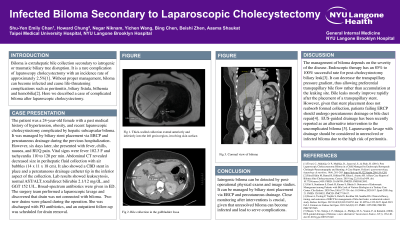
E0041 - Infected Biloma Secondary to Laparoscopic Cholecystectomy
Tuesday, October 25, 2022
3:00 PM – 5:00 PM ET
Location: Crown Ballroom

.jpg)
Shu-Yen Chan, MD, MS
Taipei Medical University Hospital
Harrisburg, Pennsylvania
Presenting Author(s)
Shu-Yen Chan, MD, MS1, Howard Chung, MD2, Negar Niknam, MD3, Yichen Wang, MD4, Bing Chen, MD5, Beishi Zheng, MD6, Aasma Shaukat, MD, MPH7
1Taipei Medical University Hospital, Harrisburg, PA; 2New York University School of Medicine, Brooklyn, NY; 3Queens Hospital Center, Queens, NY; 4Trinity Health of New England, Springfield, MA; 5New York University School of Medicine, New York, NY; 6Woodhull Medical and Mental Health Center, Brooklyn, NY; 7NYU Langone Health, New York, NY
Introduction: Biloma is an extrahepatic bile collection secondary to iatrogenic or traumatic biliary tree disruption. It is a rare complication of laparoscopy cholecystectomy with an incidence rate of approximately 2.5%. Without proper management, biloma can become infected and cause life-threatening complications such as peritonitis, biliary fistula, bilhemia and hemobilia. Here we described a case of complicated biloma after laparoscopic cholecystectomy.
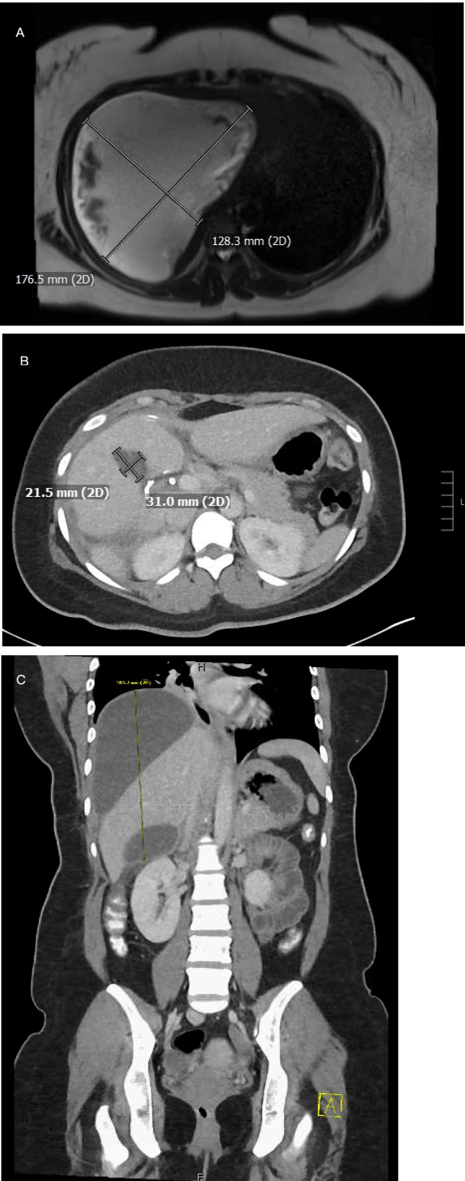
Case Description/Methods: The patient was a 24-year-old female with a past medical history of hypertension, obesity, and recent laparoscopic cholecystectomy complicated by hepatic subcapsular biloma. It was managed by biliary stent placement via endoscopic retrograde cholangiopancreatography (ERCP) and percutaneous drainage during the previous hospitalization. However, six days later, she presented with fever, chills, nausea, and right upper quadrant pain. Vital signs were fever 102.3 F and tachycardia 110 to 120 per min. The CT abdomen revealed decreased size in perihepatic fluid collection with air bubbles (14 x 11 x 18 cm). It also showed a common bile duct stent in place and a percutaneous drainage catheter tip in the inferior aspect of the collection. Lab results showed leukocytosis to 10.3, normal AST/ALT, total/direct bilirubin 2.1/12 mg/dL, and GGT 152 U/L. Broad-spectrum antibiotics were given in ED. The surgery team performed a laparoscopic lavage and discovered that the drain was not connected with the biloma. Two new drains were placed during the operation. She was discharged with PO antibiotics, and an outpatient follow-up was scheduled for drain removal.
Discussion: The management of biloma depends on the severity of the disease. Endoscopic therapy, such as a transpapillary biliary stent placement, can decrease the transpapillary pressure gradient, thus allowing preferential transpapillary bile flow rather than accumulation at the leaking site. However, given that stent placement does not reabsorb formed collection, patients failing ERCP should undergo percutaneous drainage or bile duct repair.Iatrogenic biloma can be detected by post-operational physical exams and image studies. Laparoscopic lavage with drainage should be considered in unresolved or infected biloma due to the high risk of peritonitis.

Disclosures:
Shu-Yen Chan, MD, MS1, Howard Chung, MD2, Negar Niknam, MD3, Yichen Wang, MD4, Bing Chen, MD5, Beishi Zheng, MD6, Aasma Shaukat, MD, MPH7. E0041 - Infected Biloma Secondary to Laparoscopic Cholecystectomy, ACG 2022 Annual Scientific Meeting Abstracts. Charlotte, NC: American College of Gastroenterology.
1Taipei Medical University Hospital, Harrisburg, PA; 2New York University School of Medicine, Brooklyn, NY; 3Queens Hospital Center, Queens, NY; 4Trinity Health of New England, Springfield, MA; 5New York University School of Medicine, New York, NY; 6Woodhull Medical and Mental Health Center, Brooklyn, NY; 7NYU Langone Health, New York, NY
Introduction: Biloma is an extrahepatic bile collection secondary to iatrogenic or traumatic biliary tree disruption. It is a rare complication of laparoscopy cholecystectomy with an incidence rate of approximately 2.5%. Without proper management, biloma can become infected and cause life-threatening complications such as peritonitis, biliary fistula, bilhemia and hemobilia. Here we described a case of complicated biloma after laparoscopic cholecystectomy.
Case Description/Methods: The patient was a 24-year-old female with a past medical history of hypertension, obesity, and recent laparoscopic cholecystectomy complicated by hepatic subcapsular biloma. It was managed by biliary stent placement via endoscopic retrograde cholangiopancreatography (ERCP) and percutaneous drainage during the previous hospitalization. However, six days later, she presented with fever, chills, nausea, and right upper quadrant pain. Vital signs were fever 102.3 F and tachycardia 110 to 120 per min. The CT abdomen revealed decreased size in perihepatic fluid collection with air bubbles (14 x 11 x 18 cm). It also showed a common bile duct stent in place and a percutaneous drainage catheter tip in the inferior aspect of the collection. Lab results showed leukocytosis to 10.3, normal AST/ALT, total/direct bilirubin 2.1/12 mg/dL, and GGT 152 U/L. Broad-spectrum antibiotics were given in ED. The surgery team performed a laparoscopic lavage and discovered that the drain was not connected with the biloma. Two new drains were placed during the operation. She was discharged with PO antibiotics, and an outpatient follow-up was scheduled for drain removal.
Discussion: The management of biloma depends on the severity of the disease. Endoscopic therapy, such as a transpapillary biliary stent placement, can decrease the transpapillary pressure gradient, thus allowing preferential transpapillary bile flow rather than accumulation at the leaking site. However, given that stent placement does not reabsorb formed collection, patients failing ERCP should undergo percutaneous drainage or bile duct repair.Iatrogenic biloma can be detected by post-operational physical exams and image studies. Laparoscopic lavage with drainage should be considered in unresolved or infected biloma due to the high risk of peritonitis.

Figure: A. superior dominant of subcapsular periherpatic biloma
B. collection in the gallbladder fossa
C. coronal view of the biloma
B. collection in the gallbladder fossa
C. coronal view of the biloma
Disclosures:
Shu-Yen Chan indicated no relevant financial relationships.
Howard Chung indicated no relevant financial relationships.
Negar Niknam indicated no relevant financial relationships.
Yichen Wang indicated no relevant financial relationships.
Bing Chen indicated no relevant financial relationships.
Beishi Zheng indicated no relevant financial relationships.
Aasma Shaukat indicated no relevant financial relationships.
Shu-Yen Chan, MD, MS1, Howard Chung, MD2, Negar Niknam, MD3, Yichen Wang, MD4, Bing Chen, MD5, Beishi Zheng, MD6, Aasma Shaukat, MD, MPH7. E0041 - Infected Biloma Secondary to Laparoscopic Cholecystectomy, ACG 2022 Annual Scientific Meeting Abstracts. Charlotte, NC: American College of Gastroenterology.
