Back


Poster Session C - Monday Afternoon
Category: Stomach
C0713 - A Case of Gastric Plasmablastic Lymphoma in a 30-Year-Old Immunocompetent Man
Monday, October 24, 2022
3:00 PM – 5:00 PM ET
Location: Crown Ballroom

Has Audio

Amaka Onyiagu, MD
HCA East Florida GME Westside/Northwest Internal Medicine
Plantation, FL
Presenting Author(s)
Amaka Onyiagu, MD1, Christopher O. Alabi, MD1, Mayuri Gupta, MD2
1HCA East Florida GME Westside/Northwest Internal Medicine, Plantation, FL; 2HCA Florida Northwest Hospital, Margate, FL
Introduction: Plasmablastic lymphoma (PBL) is a rare lymphoma initially predominantly seen in the oral cavity of HIV-positive individuals. Only a few cases in HIV-negative individuals and patients with no known immunocompromised status have been reported. Herein is a gastric PBL in a 30-year-old immunocompetent patient creating a diagnostic challenge.
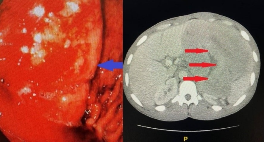
Case Description/Methods: A 30-year-old male customer care agent presented with a complaint of worsening epigastric pain. He was seen at a different hospital one month prior and diagnosed with a gastric mass with an inconclusive biopsy result. He reported nausea, hematemesis, and unintentional weight loss over three months. He denied any immunocompromised state, alcohol, and tobacco use. On physical examination, he was tachycardic with moderate epigastric tenderness. He had mild anemia with leukocytosis, positive fecal occult blood, and CT revealed a 16cm x 17cm x 16cm fundus mass in the stomach (Image 1). The Pathological and immunochemistry reports obtained after EGD (Image 1) with EUS and biopsy were in keeping with PBL. Immunodeficiency workup done was negative so was bone marrow biopsy for lymphoma. He received dose-adjusted - etoposide, prednisone, vincristine, cyclophosphamide, doxorubicin, and rituximab (da-EPOCH). He is to continue treatment courses outpatient.
Discussion: PBL is a very aggressive tumor with a median survival of 14 months and its incidence is unknown due to its rarity. A handful of case reports and small case series have been published, which stipulated the median age of diagnosis at 55 years in HIV-negative patients, with a female predominance in this group and the gastrointestinal tract as the most common tumor site (20%). PBL is difficult to diagnose because it mimics several other neoplasms. The diagnostic challenges with our patient include the initial inconclusive biopsy, the variability in his clinical presentation, and atypical immunohistochemistry findings. We are writing this to add to the available literature on cases reported that may contribute to a better understanding of this rare tumor.

Disclosures:
Amaka Onyiagu, MD1, Christopher O. Alabi, MD1, Mayuri Gupta, MD2. C0713 - A Case of Gastric Plasmablastic Lymphoma in a 30-Year-Old Immunocompetent Man, ACG 2022 Annual Scientific Meeting Abstracts. Charlotte, NC: American College of Gastroenterology.
1HCA East Florida GME Westside/Northwest Internal Medicine, Plantation, FL; 2HCA Florida Northwest Hospital, Margate, FL
Introduction: Plasmablastic lymphoma (PBL) is a rare lymphoma initially predominantly seen in the oral cavity of HIV-positive individuals. Only a few cases in HIV-negative individuals and patients with no known immunocompromised status have been reported. Herein is a gastric PBL in a 30-year-old immunocompetent patient creating a diagnostic challenge.
Case Description/Methods: A 30-year-old male customer care agent presented with a complaint of worsening epigastric pain. He was seen at a different hospital one month prior and diagnosed with a gastric mass with an inconclusive biopsy result. He reported nausea, hematemesis, and unintentional weight loss over three months. He denied any immunocompromised state, alcohol, and tobacco use. On physical examination, he was tachycardic with moderate epigastric tenderness. He had mild anemia with leukocytosis, positive fecal occult blood, and CT revealed a 16cm x 17cm x 16cm fundus mass in the stomach (Image 1). The Pathological and immunochemistry reports obtained after EGD (Image 1) with EUS and biopsy were in keeping with PBL. Immunodeficiency workup done was negative so was bone marrow biopsy for lymphoma. He received dose-adjusted - etoposide, prednisone, vincristine, cyclophosphamide, doxorubicin, and rituximab (da-EPOCH). He is to continue treatment courses outpatient.
Discussion: PBL is a very aggressive tumor with a median survival of 14 months and its incidence is unknown due to its rarity. A handful of case reports and small case series have been published, which stipulated the median age of diagnosis at 55 years in HIV-negative patients, with a female predominance in this group and the gastrointestinal tract as the most common tumor site (20%). PBL is difficult to diagnose because it mimics several other neoplasms. The diagnostic challenges with our patient include the initial inconclusive biopsy, the variability in his clinical presentation, and atypical immunohistochemistry findings. We are writing this to add to the available literature on cases reported that may contribute to a better understanding of this rare tumor.

Figure: The image on the left shows a gastric mass seen on EGD. The image on the right is a gastric mass seen on computerized tomography of the abdomen and pelvis.
Disclosures:
Amaka Onyiagu indicated no relevant financial relationships.
Christopher Alabi indicated no relevant financial relationships.
Mayuri Gupta indicated no relevant financial relationships.
Amaka Onyiagu, MD1, Christopher O. Alabi, MD1, Mayuri Gupta, MD2. C0713 - A Case of Gastric Plasmablastic Lymphoma in a 30-Year-Old Immunocompetent Man, ACG 2022 Annual Scientific Meeting Abstracts. Charlotte, NC: American College of Gastroenterology.
