Back


Poster Session C - Monday Afternoon
Category: General Endoscopy
C0298 - Gastrosplenic Fistula - An Open and Closed Case
Monday, October 24, 2022
3:00 PM – 5:00 PM ET
Location: Crown Ballroom

Has Audio
- TM
Thomas J. Mathews, MD
University of Kansas Medical Center
Kansas City, KS
Presenting Author(s)
Thomas J. Mathews, MD, Mitesh Patel, MD
University of Kansas Medical Center, Kansas City, KS
Introduction: In this case, we will discuss endoscopic treatment for immunocompromised patients with gastrosplenic fistulas.
Case Description/Methods: A 36-year-old Caucasian female with history of diffuse large B cell lymphoma (DLBCL) on rituximab, cyclophosphamide, doxorubicin, vincristine, and prednisone (R-CHOP) chemotherapy presented with neutropenic fever, persistent cough with deep inspiration, and burning pain in her left shoulder that had worsened since chemotherapy two weeks prior.
On admission, she was tachycardic and afebrile. White blood cell count was 1,200/mm3, absolute neutrophil count 120/mm3, and blood cultures were negative. CT demonstrated improved splenomegaly measuring 6.9 x 7.0 cm (previously 13 x 11 cm) and moderate gas and fluid within the spleen with suspected fistulous communication to the adjacent greater curvature of the stomach. An upper GI series confirmed gastrosplenic fistula (GSF) and fluid collection in the spleen. Gastroenterology and interventional radiology (IR) were consulted.
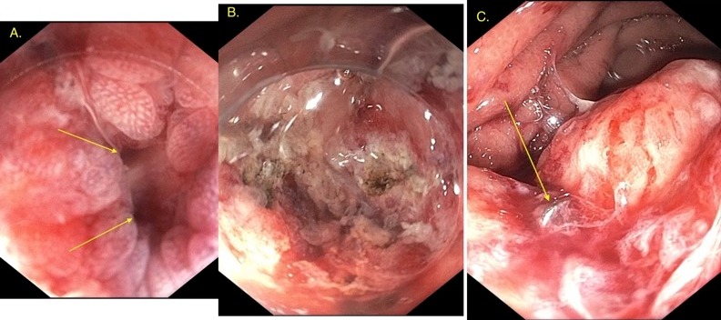
Endoscopy revealed diffuse, severely congested mucosa in the gastric fundus and body, causing difficulty visualizing the fistula. An endocap was attached to the endoscope to assist with visualization. A 5 mm fistula with ulceration was found on the greater curvature of the gastric body. Argon plasma coagulation was performed for tissue devitalization in and around the fistula. The scope was then outfitted with an over-the-scope clip, which successfully closed the fistula.
IR then placed an abdominal drain, and fluid cultures grew Streptococcus constellatus, Streptococcus anginosus, Lactobacillus rhamnosus, and Parvimonas micra. Empiric antibiotics were changed to intravenous ertapenem. The patient was able to tolerate a regular diet. Three weeks later, CT abdomen revealed significant decrease in size of splenic gas and fluid collections.
Discussion: GSFs are a rare complication in patients with lymphoma and occur almost exclusively in patients with DLBCL. This patient did not present with typical features such as melena, hematochezia, severe sepsis, severe abdominal pain, nausea, vomiting, or hematemesis. It is important to consider GSF in patients with DLBCL presenting with GI issues.
The overwhelming majority of GSF cases are surgical emergencies, but not every case will require surgical intervention. This can be important in patients at increased risk of surgical morbidity and mortality. A GI consult could save patients from unnecessary risk and financial burden.

Disclosures:
Thomas J. Mathews, MD, Mitesh Patel, MD. C0298 - Gastrosplenic Fistula - An Open and Closed Case, ACG 2022 Annual Scientific Meeting Abstracts. Charlotte, NC: American College of Gastroenterology.
University of Kansas Medical Center, Kansas City, KS
Introduction: In this case, we will discuss endoscopic treatment for immunocompromised patients with gastrosplenic fistulas.
Case Description/Methods: A 36-year-old Caucasian female with history of diffuse large B cell lymphoma (DLBCL) on rituximab, cyclophosphamide, doxorubicin, vincristine, and prednisone (R-CHOP) chemotherapy presented with neutropenic fever, persistent cough with deep inspiration, and burning pain in her left shoulder that had worsened since chemotherapy two weeks prior.
On admission, she was tachycardic and afebrile. White blood cell count was 1,200/mm3, absolute neutrophil count 120/mm3, and blood cultures were negative. CT demonstrated improved splenomegaly measuring 6.9 x 7.0 cm (previously 13 x 11 cm) and moderate gas and fluid within the spleen with suspected fistulous communication to the adjacent greater curvature of the stomach. An upper GI series confirmed gastrosplenic fistula (GSF) and fluid collection in the spleen. Gastroenterology and interventional radiology (IR) were consulted.
Endoscopy revealed diffuse, severely congested mucosa in the gastric fundus and body, causing difficulty visualizing the fistula. An endocap was attached to the endoscope to assist with visualization. A 5 mm fistula with ulceration was found on the greater curvature of the gastric body. Argon plasma coagulation was performed for tissue devitalization in and around the fistula. The scope was then outfitted with an over-the-scope clip, which successfully closed the fistula.
IR then placed an abdominal drain, and fluid cultures grew Streptococcus constellatus, Streptococcus anginosus, Lactobacillus rhamnosus, and Parvimonas micra. Empiric antibiotics were changed to intravenous ertapenem. The patient was able to tolerate a regular diet. Three weeks later, CT abdomen revealed significant decrease in size of splenic gas and fluid collections.
Discussion: GSFs are a rare complication in patients with lymphoma and occur almost exclusively in patients with DLBCL. This patient did not present with typical features such as melena, hematochezia, severe sepsis, severe abdominal pain, nausea, vomiting, or hematemesis. It is important to consider GSF in patients with DLBCL presenting with GI issues.
The overwhelming majority of GSF cases are surgical emergencies, but not every case will require surgical intervention. This can be important in patients at increased risk of surgical morbidity and mortality. A GI consult could save patients from unnecessary risk and financial burden.

Figure: A. Gastrosplenic fistula (gastric body) B. After APC C. After Clip Deployment
Disclosures:
Thomas Mathews indicated no relevant financial relationships.
Mitesh Patel indicated no relevant financial relationships.
Thomas J. Mathews, MD, Mitesh Patel, MD. C0298 - Gastrosplenic Fistula - An Open and Closed Case, ACG 2022 Annual Scientific Meeting Abstracts. Charlotte, NC: American College of Gastroenterology.
