Back


Poster Session C - Monday Afternoon
Category: General Endoscopy
C0303 - Breast Cancer With Synchronous Endoluminal Upper and Lower GI Metastases
Monday, October 24, 2022
3:00 PM – 5:00 PM ET
Location: Crown Ballroom

Has Audio

Tiberiu Moga, MD
University at Buffalo
Buffalo, NY
Presenting Author(s)
Tiberiu Moga, MD1, Kevin Robillard, MD2
1University at Buffalo, Buffalo, NY; 2Roswell Park Comprehensive Cancer Center, Buffalo, NY
Introduction: Breast cancer is the most common cancer of women and spreads commonly to organs such as the lungs, bones, brain and liver. We present a case of a 61 year-old woman who had isolated upper and lower GI endoluminal metastases from a primary breast lobular carcinoma in situ (LCIS) lesion.
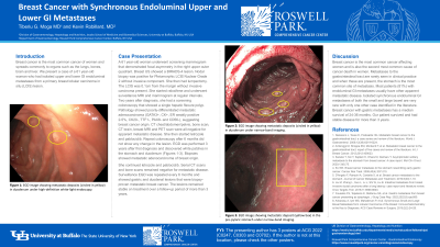
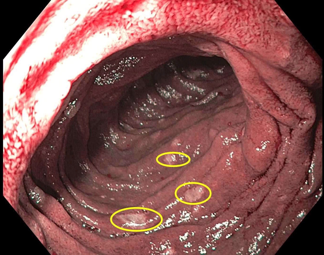
Case Description/Methods: A 61 year-old woman underwent screening mammogram that demonstrated focal asymmetry in the right upper outer quadrant. Breast US showed a BIRADS-4 lesion. Nodal biopsy was positive for Pleiomorphic LCIS Nuclear Grade 2 without invasive component. She then had lumpectomy. The LCIS was 0.1cm from the margin without invasive carcinoma present. She started raloxifene and underwent surveillance MRI and mammogram at regular intervals. Two years after diagnosis, she had a screening colonoscopy that showed a single hepatic flexure polyp. Pathology showed poorly-differentiated metastatic adenocarcinoma (GATA3+, CK+, ER weakly positive 2-5%, CK20-, TTF1-, PAX8- and CD56-), suggesting breast cancer origin. CT chest/abdomen/pelvis, bone scan, CT brain, breast MRI and PET scan were all negative for apparent metastatic disease. She then started letrozole and pablociclib. Repeat colonoscopy after 6 months did not show any change in the lesion. EGD was performed 3 years after first diagnosis and discovered white patches in the duodenum. Biopsies showed metastatic adenocarcinoma of breast origin.
She continued letrozole and pablociclib. Serial CT scans and bone scans remained negative for metastatic disease. Surveillance EGD was repeated every 6 months and showed gastric and duodenal lesions that were biopsy-proven metastatic breast cancer. The lesions remained stable on treatment over a follow-up period of more than 3 years.
Discussion: Breast cancer is the most common cancer affecting women and is also the second most common cause of cancer death in women. Metastases to the gastrointestinal tract are rarely seen in clinical practice and when these are present, the stomach is the most common site of metastasis. Most patients (81%) with endoluminal GI metastases usually have other apparent metastatic disease. Isolated synchronous endoluminal GI metastases of both the small and large bowel are very rare with only one other case identified in the literature. Breast cancer with gastric metastases has a median survival of 24-36 months. Our patient survived and had stable disease for more than 3 years.

Disclosures:
Tiberiu Moga, MD1, Kevin Robillard, MD2. C0303 - Breast Cancer With Synchronous Endoluminal Upper and Lower GI Metastases, ACG 2022 Annual Scientific Meeting Abstracts. Charlotte, NC: American College of Gastroenterology.
1University at Buffalo, Buffalo, NY; 2Roswell Park Comprehensive Cancer Center, Buffalo, NY
Introduction: Breast cancer is the most common cancer of women and spreads commonly to organs such as the lungs, bones, brain and liver. We present a case of a 61 year-old woman who had isolated upper and lower GI endoluminal metastases from a primary breast lobular carcinoma in situ (LCIS) lesion.
Case Description/Methods: A 61 year-old woman underwent screening mammogram that demonstrated focal asymmetry in the right upper outer quadrant. Breast US showed a BIRADS-4 lesion. Nodal biopsy was positive for Pleiomorphic LCIS Nuclear Grade 2 without invasive component. She then had lumpectomy. The LCIS was 0.1cm from the margin without invasive carcinoma present. She started raloxifene and underwent surveillance MRI and mammogram at regular intervals. Two years after diagnosis, she had a screening colonoscopy that showed a single hepatic flexure polyp. Pathology showed poorly-differentiated metastatic adenocarcinoma (GATA3+, CK+, ER weakly positive 2-5%, CK20-, TTF1-, PAX8- and CD56-), suggesting breast cancer origin. CT chest/abdomen/pelvis, bone scan, CT brain, breast MRI and PET scan were all negative for apparent metastatic disease. She then started letrozole and pablociclib. Repeat colonoscopy after 6 months did not show any change in the lesion. EGD was performed 3 years after first diagnosis and discovered white patches in the duodenum. Biopsies showed metastatic adenocarcinoma of breast origin.
She continued letrozole and pablociclib. Serial CT scans and bone scans remained negative for metastatic disease. Surveillance EGD was repeated every 6 months and showed gastric and duodenal lesions that were biopsy-proven metastatic breast cancer. The lesions remained stable on treatment over a follow-up period of more than 3 years.
Discussion: Breast cancer is the most common cancer affecting women and is also the second most common cause of cancer death in women. Metastases to the gastrointestinal tract are rarely seen in clinical practice and when these are present, the stomach is the most common site of metastasis. Most patients (81%) with endoluminal GI metastases usually have other apparent metastatic disease. Isolated synchronous endoluminal GI metastases of both the small and large bowel are very rare with only one other case identified in the literature. Breast cancer with gastric metastases has a median survival of 24-36 months. Our patient survived and had stable disease for more than 3 years.

Figure: Figure 1: Duodenum examined using Narrow-Band Imaging (NBI) showing metastatic deposits (circled in yellow).
Disclosures:
Tiberiu Moga indicated no relevant financial relationships.
Kevin Robillard indicated no relevant financial relationships.
Tiberiu Moga, MD1, Kevin Robillard, MD2. C0303 - Breast Cancer With Synchronous Endoluminal Upper and Lower GI Metastases, ACG 2022 Annual Scientific Meeting Abstracts. Charlotte, NC: American College of Gastroenterology.
