Back


Poster Session C - Monday Afternoon
Category: General Endoscopy
C0309 - Clinically Significant Upper Gastrointestinal Bleeding Post Esophagogastroduodenoscopy Cold Biopsy: A Rare Case Report
Monday, October 24, 2022
3:00 PM – 5:00 PM ET
Location: Crown Ballroom

Has Audio

Michelle D'Souza, MD
University of Rochester Medical Center
Rochester, NY
Presenting Author(s)
Michelle D'Souza, MD, Abigail Schubach, MD, Matthew Berger, MD, Andrew Brown, MD, Vivek Kaul, MD, FACG
University of Rochester Medical Center, Rochester, NY
Introduction: Bleeding after cold forceps biopsy of the gastrointestinal tract is an extremely rare phenomenon, estimated incidence < 0.1%. Clinically significant bleeding is even rarer. However, in patients who have evidence of gastrointestinal bleeding (GIB) after endoscopic biopsy, it is an important cause to consider.
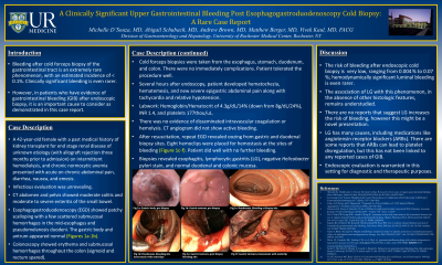
Case Description/Methods: A 42 year old female with a past medical history of kidney transplant for end stage renal disease of unknown etiology (with allograft rejection three months prior to admission), on intermittent hemodialysis and chronic normocytic anemia presented with acute on chronic abdominal pain, diarrhea, nausea, and emesis. Infectious evaluation was unrevealing. CT abdomen and pelvis showed moderate colitis and moderate to severe enteritis of the mid small bowel. Esophagogastroduodenoscopy (EGD) showed scalloping of the mucosa with a few scattered submucosal hemorrhages in the mid-esophagus and pseudomelanosis duodeni. Colonoscopy showed erythema and submucosal hemorrhages throughout the colon (sigmoid and rectum spared). Cold forceps biopsies were taken from the esophagus, stomach, duodenum, and colon.
Several hours after endoscopy, she had hematochezia, hematemesis, and new severe epigastric abdominal pain along with tachycardia and relative hypotension. Labwork revealed a hemoglobin/hematocrit of 4.3g/dL/14% (down from 8g/dL/24%), INR 1.4, and platelets 177thou/uL. There was no evidence of disseminated intravascular coagulation or hemolysis. CT angiogram did not show active bleeding. After resuscitation, repeat EGD revealed oozing from gastric and duodenal biopsy sites. Eight hemoclips were placed for hemostasis at the sites of bleeding. Biopsies from initial EGD revealed esophagitis, lymphocytic gastritis (LG), negative helicobacter pylori stain, and normal duodenal and colonic mucosa.
Discussion: The risk of bleeding after endoscopic cold biopsy is rare, ranging from 0.004 % to 0.07 %; hemodynamically significant luminal bleeding is even rarer. The association of LG with this phenomenon, in the absence of other histologic features, remains understudied. There are no reports that suggest LG increases the risk of bleeding, however this might be a novel presentation. LG has many causes, including medications like angiotensin receptor blockers (ARBs). There are some reports that ARBs can lead to platelet disregulation, but this has not be linked to any cases of GIB. Endoscopic evaluation is warranted in this setting for diagnostic and therapeutic purposes.

Disclosures:
Michelle D'Souza, MD, Abigail Schubach, MD, Matthew Berger, MD, Andrew Brown, MD, Vivek Kaul, MD, FACG. C0309 - Clinically Significant Upper Gastrointestinal Bleeding Post Esophagogastroduodenoscopy Cold Biopsy: A Rare Case Report, ACG 2022 Annual Scientific Meeting Abstracts. Charlotte, NC: American College of Gastroenterology.
University of Rochester Medical Center, Rochester, NY
Introduction: Bleeding after cold forceps biopsy of the gastrointestinal tract is an extremely rare phenomenon, estimated incidence < 0.1%. Clinically significant bleeding is even rarer. However, in patients who have evidence of gastrointestinal bleeding (GIB) after endoscopic biopsy, it is an important cause to consider.
Case Description/Methods: A 42 year old female with a past medical history of kidney transplant for end stage renal disease of unknown etiology (with allograft rejection three months prior to admission), on intermittent hemodialysis and chronic normocytic anemia presented with acute on chronic abdominal pain, diarrhea, nausea, and emesis. Infectious evaluation was unrevealing. CT abdomen and pelvis showed moderate colitis and moderate to severe enteritis of the mid small bowel. Esophagogastroduodenoscopy (EGD) showed scalloping of the mucosa with a few scattered submucosal hemorrhages in the mid-esophagus and pseudomelanosis duodeni. Colonoscopy showed erythema and submucosal hemorrhages throughout the colon (sigmoid and rectum spared). Cold forceps biopsies were taken from the esophagus, stomach, duodenum, and colon.
Several hours after endoscopy, she had hematochezia, hematemesis, and new severe epigastric abdominal pain along with tachycardia and relative hypotension. Labwork revealed a hemoglobin/hematocrit of 4.3g/dL/14% (down from 8g/dL/24%), INR 1.4, and platelets 177thou/uL. There was no evidence of disseminated intravascular coagulation or hemolysis. CT angiogram did not show active bleeding. After resuscitation, repeat EGD revealed oozing from gastric and duodenal biopsy sites. Eight hemoclips were placed for hemostasis at the sites of bleeding. Biopsies from initial EGD revealed esophagitis, lymphocytic gastritis (LG), negative helicobacter pylori stain, and normal duodenal and colonic mucosa.
Discussion: The risk of bleeding after endoscopic cold biopsy is rare, ranging from 0.004 % to 0.07 %; hemodynamically significant luminal bleeding is even rarer. The association of LG with this phenomenon, in the absence of other histologic features, remains understudied. There are no reports that suggest LG increases the risk of bleeding, however this might be a novel presentation. LG has many causes, including medications like angiotensin receptor blockers (ARBs). There are some reports that ARBs can lead to platelet disregulation, but this has not be linked to any cases of GIB. Endoscopic evaluation is warranted in this setting for diagnostic and therapeutic purposes.

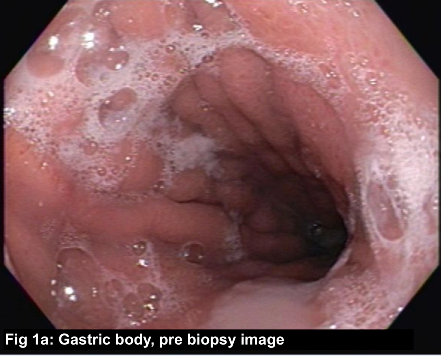
Figure: Figure 1: Pre and Post Biopsy Endoscopic Images
Disclosures:
Michelle D'Souza indicated no relevant financial relationships.
Abigail Schubach indicated no relevant financial relationships.
Matthew Berger indicated no relevant financial relationships.
Andrew Brown indicated no relevant financial relationships.
Vivek Kaul: Ambu – Consultant. CDX – Consultant. Cook Medical – Consultant. Motus GI – Consultant. Steris Corp – Consultant.
Michelle D'Souza, MD, Abigail Schubach, MD, Matthew Berger, MD, Andrew Brown, MD, Vivek Kaul, MD, FACG. C0309 - Clinically Significant Upper Gastrointestinal Bleeding Post Esophagogastroduodenoscopy Cold Biopsy: A Rare Case Report, ACG 2022 Annual Scientific Meeting Abstracts. Charlotte, NC: American College of Gastroenterology.
