Back


Poster Session A - Sunday Afternoon
Category: Liver
A0556 - Vanishing Bile Duct Syndrome in the Setting of Newly Diagnosed Hodgkin Lymphoma
Sunday, October 23, 2022
5:00 PM – 7:00 PM ET
Location: Crown Ballroom

Has Audio

Abdullah A. Muftah, MD
Baylor College of Medicine
Houston, Texas
Presenting Author(s)
Abdullah A. Muftah, MD1, Rebecca Waters, DO2, Lan Wang, MD3, Hao Chi Zhang, MD4
1Baylor College of Medicine, Houston, TX; 2University of Texas MD Anderson Cancer Center, Houston, TX; 3MD Anderson Cancer Center, Houston, TX; 4UT MD Anderson Cancer Center, Houston, TX
Introduction: Vanishing bile duct syndrome (VBDS) is characterized by cholestatic liver disease in the setting of disappearing intra-hepatic bile ducts. It can resemble other forms of cholestatic liver disease; however, imaging and biochemical tests will be unrevealing. We describe a case of a woman with Hodgkin lymphoma (HL) who developed cholestatic hepatitis with loss of intra-hepatic bile ducts.
Case Description/Methods: A 23-year-old woman with a history of HL who presented for evaluation and treatment of HL. Two months prior she had been treated for CMV-associated gastroenteritis and sepsis. Initial examination revealed jaundice and bilateral conjunctival icterus.. Lab results showed total bilirubin of 18.1 mg/dL, ALT 363 U/L, AST 149 U/L, alkaline phosphatase (ALP) of 2392 U/L, direct bilirubin of 15.5 mg/dL, INR of 2.07, and cytomegalovirus (CMV) PCR at 670 IU/mL. Other acute viral hepatitis studies, tests for intrinsic liver disease, and autoimmune markers were negative. Computerized tomography (CT) of the abdomen and pelvis showed extensive lymphadenopathy, sclerotic changes throughout the skeleton, splenomegaly, and hepatomegaly without focal liver lesions. Magnetic resonance cholangiopancreatography (MRCP) of the abdomen showed no evidence of biliary ductal dilatation. Treatment for her HL was initiated, as well as foscarnet for her CMV viremia. Liver biopsy showed benign liver parenchyma with marked cholestasis and paucity of bile ducts, no cirrhosis, and no viral inclusions. The patient was initiated on ursodeoxycholic acid (UDCA) at 15 mg/kg/d without resolution of symptoms or cholestasis. Given no improvement in her cholestasis, the prospects of liver transplantation (LT) were discussed, but the patient was not a candidate.
Discussion: Liver involvement in HL typically manifests as parenchymal invasion, external compression, or paraneoplastic destruction of bile ducts (VBDS). VBDS tends to signify a poor prognosis with patients frequently progressing to liver failure. The pathogenesis of VBDS in HL is not yet defined with current evidence suggesting an immune-mediated response. Typically, viral causes should be excluded. In this case, the patient did have CMV viremia. Given her prior treatment, low viral load, and absence of viral inclusions on liver biopsy, CMV was excluded as a cause. Treatment for VBDS revolves around treating the underlying cause. UDCA can be used as a temporizing measure, but, if no improvement is observed over time, patients are referred for liver transplant.

Disclosures:
Abdullah A. Muftah, MD1, Rebecca Waters, DO2, Lan Wang, MD3, Hao Chi Zhang, MD4. A0556 - Vanishing Bile Duct Syndrome in the Setting of Newly Diagnosed Hodgkin Lymphoma, ACG 2022 Annual Scientific Meeting Abstracts. Charlotte, NC: American College of Gastroenterology.
1Baylor College of Medicine, Houston, TX; 2University of Texas MD Anderson Cancer Center, Houston, TX; 3MD Anderson Cancer Center, Houston, TX; 4UT MD Anderson Cancer Center, Houston, TX
Introduction: Vanishing bile duct syndrome (VBDS) is characterized by cholestatic liver disease in the setting of disappearing intra-hepatic bile ducts. It can resemble other forms of cholestatic liver disease; however, imaging and biochemical tests will be unrevealing. We describe a case of a woman with Hodgkin lymphoma (HL) who developed cholestatic hepatitis with loss of intra-hepatic bile ducts.
Case Description/Methods: A 23-year-old woman with a history of HL who presented for evaluation and treatment of HL. Two months prior she had been treated for CMV-associated gastroenteritis and sepsis. Initial examination revealed jaundice and bilateral conjunctival icterus.. Lab results showed total bilirubin of 18.1 mg/dL, ALT 363 U/L, AST 149 U/L, alkaline phosphatase (ALP) of 2392 U/L, direct bilirubin of 15.5 mg/dL, INR of 2.07, and cytomegalovirus (CMV) PCR at 670 IU/mL. Other acute viral hepatitis studies, tests for intrinsic liver disease, and autoimmune markers were negative. Computerized tomography (CT) of the abdomen and pelvis showed extensive lymphadenopathy, sclerotic changes throughout the skeleton, splenomegaly, and hepatomegaly without focal liver lesions. Magnetic resonance cholangiopancreatography (MRCP) of the abdomen showed no evidence of biliary ductal dilatation. Treatment for her HL was initiated, as well as foscarnet for her CMV viremia. Liver biopsy showed benign liver parenchyma with marked cholestasis and paucity of bile ducts, no cirrhosis, and no viral inclusions. The patient was initiated on ursodeoxycholic acid (UDCA) at 15 mg/kg/d without resolution of symptoms or cholestasis. Given no improvement in her cholestasis, the prospects of liver transplantation (LT) were discussed, but the patient was not a candidate.
Discussion: Liver involvement in HL typically manifests as parenchymal invasion, external compression, or paraneoplastic destruction of bile ducts (VBDS). VBDS tends to signify a poor prognosis with patients frequently progressing to liver failure. The pathogenesis of VBDS in HL is not yet defined with current evidence suggesting an immune-mediated response. Typically, viral causes should be excluded. In this case, the patient did have CMV viremia. Given her prior treatment, low viral load, and absence of viral inclusions on liver biopsy, CMV was excluded as a cause. Treatment for VBDS revolves around treating the underlying cause. UDCA can be used as a temporizing measure, but, if no improvement is observed over time, patients are referred for liver transplant.

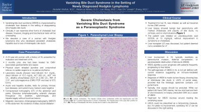
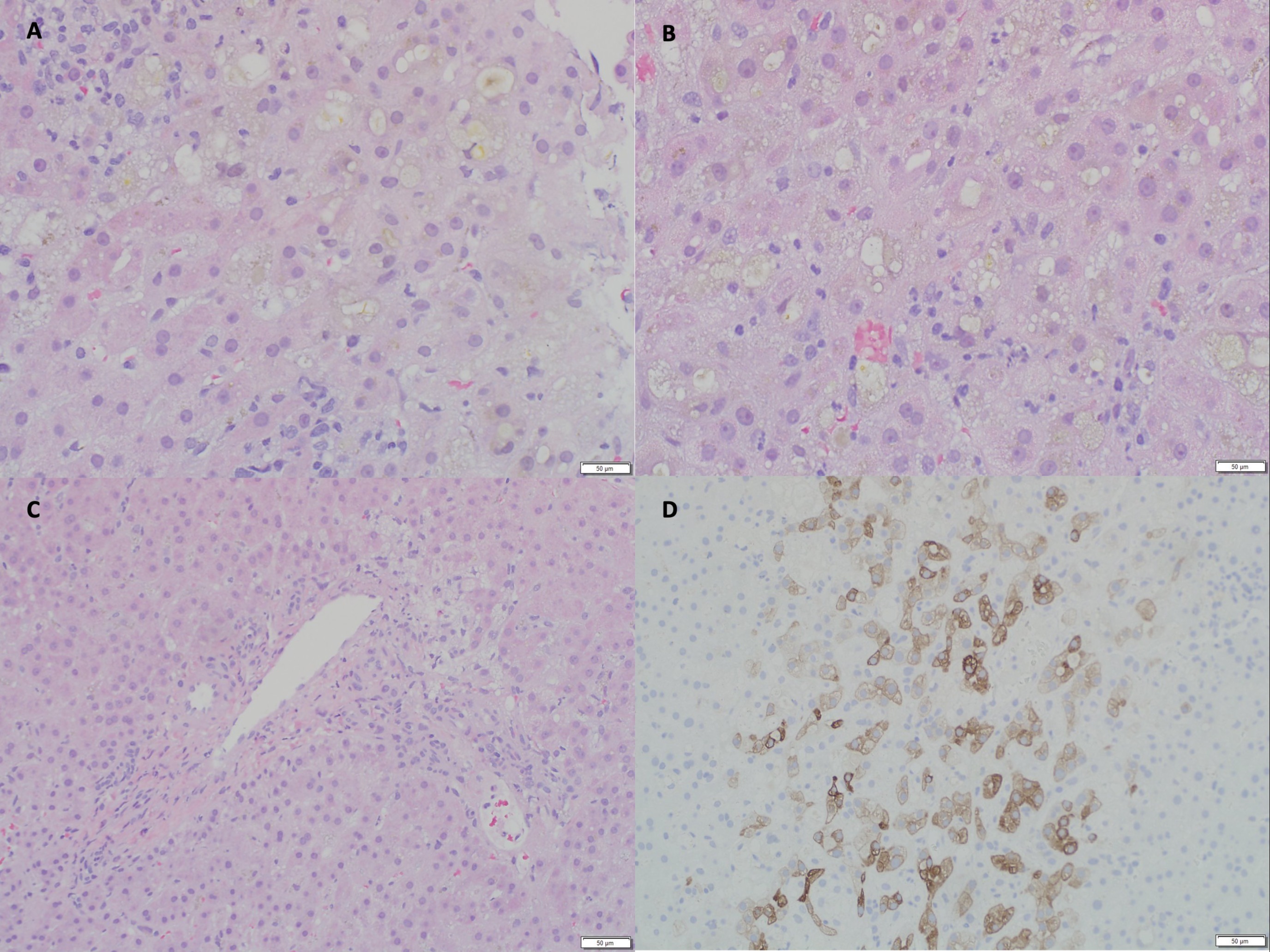
Figure: Parenchymal liver biopsy: (A) and (B): Lobular cholestasis. The hepatic lobules show cholestasis within the hepatocytes and bile canaliculi. (C): Paucity of intrahepatic bile duct. No bile duct is seen in the portal tract. (D): CK-7 positive hepatocytes in chronic cholestasis. The hepatocytes in this case stain positive for CK-7.
Disclosures:
Abdullah Muftah indicated no relevant financial relationships.
Rebecca Waters: Astellas – Advisor or Review Panel Member.
Lan Wang indicated no relevant financial relationships.
Hao Chi Zhang indicated no relevant financial relationships.
Abdullah A. Muftah, MD1, Rebecca Waters, DO2, Lan Wang, MD3, Hao Chi Zhang, MD4. A0556 - Vanishing Bile Duct Syndrome in the Setting of Newly Diagnosed Hodgkin Lymphoma, ACG 2022 Annual Scientific Meeting Abstracts. Charlotte, NC: American College of Gastroenterology.
