Back


Poster Session A - Sunday Afternoon
Category: Colon
A0112 - Case of Ileocolonic Intussusception With Tubulovillous and Tubular Adenoma Lead Points in a 43-Year-Old Male
Sunday, October 23, 2022
5:00 PM – 7:00 PM ET
Location: Crown Ballroom

Has Audio

Mudassar K. Sandozi, DO
MercyHealth Internal Medicine Residency
Rockford, IL
Presenting Author(s)
Mudassar K. Sandozi, DO1, Janine B. Borja, MD2, Altaf Dawood, MD3, Naser Khan, MD2, Ammar Aqeel, MD4, Luqman Baloch, MD3
1MercyHealth Internal Medicine Residency, Rockford, IL; 2MercyHealth, Rockford, IL; 3MercyHealth Internal Medicine Residency Program, Rockford, IL; 4MercyHealth System, Rockford, IL
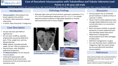
Introduction: Intussusception describes the telescoping of one segment of bowel into another. Though idiopathic intussusception most commonly causes bowel obstruction in children, intussusception in adults is uncommon and suggests malignancy. We present a case where intussusception symptoms allowed for the identification and resection of precancerous lesions in a patient.
Case Description/Methods: A 43-year-old male with history of hypertension presented with one-year history of diffuse, episodic, progressively worsening abdominal pain, 75 lbs. weight loss, and non-bloody diarrhea. He presented afebrile, hemodynamically stable, saturating well on room air. He displayed mild, right sided abdominal tenderness and CT scan showed 43 mm ileocolic intussusception. Patient underwent laparoscopic right hemicolectomy with end-to-end anastomosis of the terminal ileum and transverse colon. Resected right colon and terminal ileum specimen contained a 5.2x4.5x4.0-cm tubulovillous adenoma and 2.2x2.2x1.7-cm tubular adenoma without evidence of high-grade dysplasia or invasive adenocarcinoma. Resected colon tissue margins were free from adenomas. Surgical recovery and discharge progressed without incident.
Discussion: Intussusception rarely occurs in adults, with an annual incidence of ~2-3 cases per 1 million. Though classically associated with the triad of colicky abdominal pain, sausage-shaped abdominal mass, and currant jelly stools, in adults, intussusception commonly manifests as episodic paroxysms of severe abdominal pain and symptoms of bowel obstruction. Abdomen CT can distinguish intussusception from other etiologies of obstruction. Physicians classify intussusception by location, natural progression (fixed vs intermittent), and presence of lead point. Malignancy most frequently serves as the lead point in adults. Definitive management is surgery. Our patient presented with intermittent abdominal pain and weight loss. Ileocolonic intussusception was identified on CT, and patient underwent resection of affected bowel. Cecal tubulovillous and tubular adenomas, both > 2 cm in size, served as lead points and showed no signs of dysplasia. Intussusception of these lesions alerted clinicians to their presence so they could be resected prior to becoming dysplastic and cancerous.

Disclosures:
Mudassar K. Sandozi, DO1, Janine B. Borja, MD2, Altaf Dawood, MD3, Naser Khan, MD2, Ammar Aqeel, MD4, Luqman Baloch, MD3. A0112 - Case of Ileocolonic Intussusception With Tubulovillous and Tubular Adenoma Lead Points in a 43-Year-Old Male, ACG 2022 Annual Scientific Meeting Abstracts. Charlotte, NC: American College of Gastroenterology.
1MercyHealth Internal Medicine Residency, Rockford, IL; 2MercyHealth, Rockford, IL; 3MercyHealth Internal Medicine Residency Program, Rockford, IL; 4MercyHealth System, Rockford, IL
Introduction: Intussusception describes the telescoping of one segment of bowel into another. Though idiopathic intussusception most commonly causes bowel obstruction in children, intussusception in adults is uncommon and suggests malignancy. We present a case where intussusception symptoms allowed for the identification and resection of precancerous lesions in a patient.
Case Description/Methods: A 43-year-old male with history of hypertension presented with one-year history of diffuse, episodic, progressively worsening abdominal pain, 75 lbs. weight loss, and non-bloody diarrhea. He presented afebrile, hemodynamically stable, saturating well on room air. He displayed mild, right sided abdominal tenderness and CT scan showed 43 mm ileocolic intussusception. Patient underwent laparoscopic right hemicolectomy with end-to-end anastomosis of the terminal ileum and transverse colon. Resected right colon and terminal ileum specimen contained a 5.2x4.5x4.0-cm tubulovillous adenoma and 2.2x2.2x1.7-cm tubular adenoma without evidence of high-grade dysplasia or invasive adenocarcinoma. Resected colon tissue margins were free from adenomas. Surgical recovery and discharge progressed without incident.
Discussion: Intussusception rarely occurs in adults, with an annual incidence of ~2-3 cases per 1 million. Though classically associated with the triad of colicky abdominal pain, sausage-shaped abdominal mass, and currant jelly stools, in adults, intussusception commonly manifests as episodic paroxysms of severe abdominal pain and symptoms of bowel obstruction. Abdomen CT can distinguish intussusception from other etiologies of obstruction. Physicians classify intussusception by location, natural progression (fixed vs intermittent), and presence of lead point. Malignancy most frequently serves as the lead point in adults. Definitive management is surgery. Our patient presented with intermittent abdominal pain and weight loss. Ileocolonic intussusception was identified on CT, and patient underwent resection of affected bowel. Cecal tubulovillous and tubular adenomas, both > 2 cm in size, served as lead points and showed no signs of dysplasia. Intussusception of these lesions alerted clinicians to their presence so they could be resected prior to becoming dysplastic and cancerous.

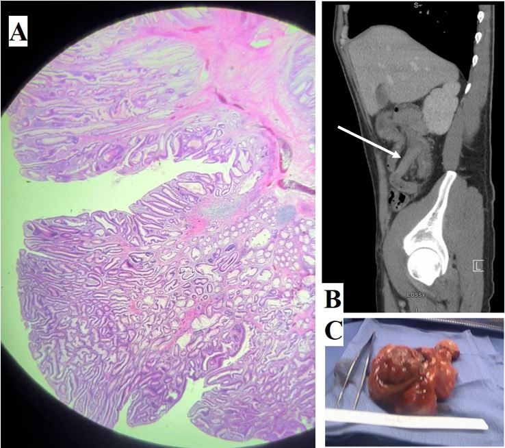
Figure: A – Characteristic tubulovillous adenoma on path.
B - Saggital view showing lead point mass, intussceptum and invaginated mesentery in right upper quadrant just inferior to the liver border and indicated by arrow.
C - Resected segment of cecum found to have 5.2 x 4.5 x 4.0 cm tubulovillous adenoma and 2.2 x 2.2 x 1.7 cm tubular adenoma, the presumed lead points of intussusception.
Disclosures:
Mudassar Sandozi indicated no relevant financial relationships.
Janine Borja indicated no relevant financial relationships.
Altaf Dawood indicated no relevant financial relationships.
Naser Khan indicated no relevant financial relationships.
Ammar Aqeel indicated no relevant financial relationships.
Luqman Baloch indicated no relevant financial relationships.
Mudassar K. Sandozi, DO1, Janine B. Borja, MD2, Altaf Dawood, MD3, Naser Khan, MD2, Ammar Aqeel, MD4, Luqman Baloch, MD3. A0112 - Case of Ileocolonic Intussusception With Tubulovillous and Tubular Adenoma Lead Points in a 43-Year-Old Male, ACG 2022 Annual Scientific Meeting Abstracts. Charlotte, NC: American College of Gastroenterology.
