Back


Poster Session A - Sunday Afternoon
Category: Colon
A0120 - Primary Colonic B-Cell Lymphoma in a Young Patient
Sunday, October 23, 2022
5:00 PM – 7:00 PM ET
Location: Crown Ballroom

Has Audio

Hassan Zreik, MD
Henry Ford Jackson
Dearborn, MI
Presenting Author(s)
Hassan Zreik, MD, Parth M. Patel, MD, Sruthi Ramanan, MD, Harjinder Singh, MD, Shamik Parikh, MD, Merritt Bern, MD
Henry Ford Jackson, Jackson, MI
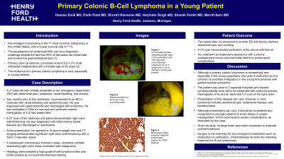
Introduction: The development of extranodal NHL can be a diagnostic challenge despite the fact that 30% of the cases are extranodal and involve the gastrointestinal tract. The incidence of a primary colonic lymphoma is rare, especially in young patients.
Case Description/Methods: A 21-year-old man initially presented to our emergency department (ED) with abdominal pain, weakness, rectal bleeding, and anemia. Two months prior to this admission, he presented to an ED in Colorado with rectal bleeding and abdominal pain. He was diagnosed with gastroenteritis and discharged with antibiotics. He was evaluated in Ohio for syncope and profound anemia (hemoglobin of 4.2) two weeks later. A CT scan of the abdomen and pelvis demonstrated right colon wall thickening. He was diagnosed with inflammatory bowel disease and discharged on prednisone.
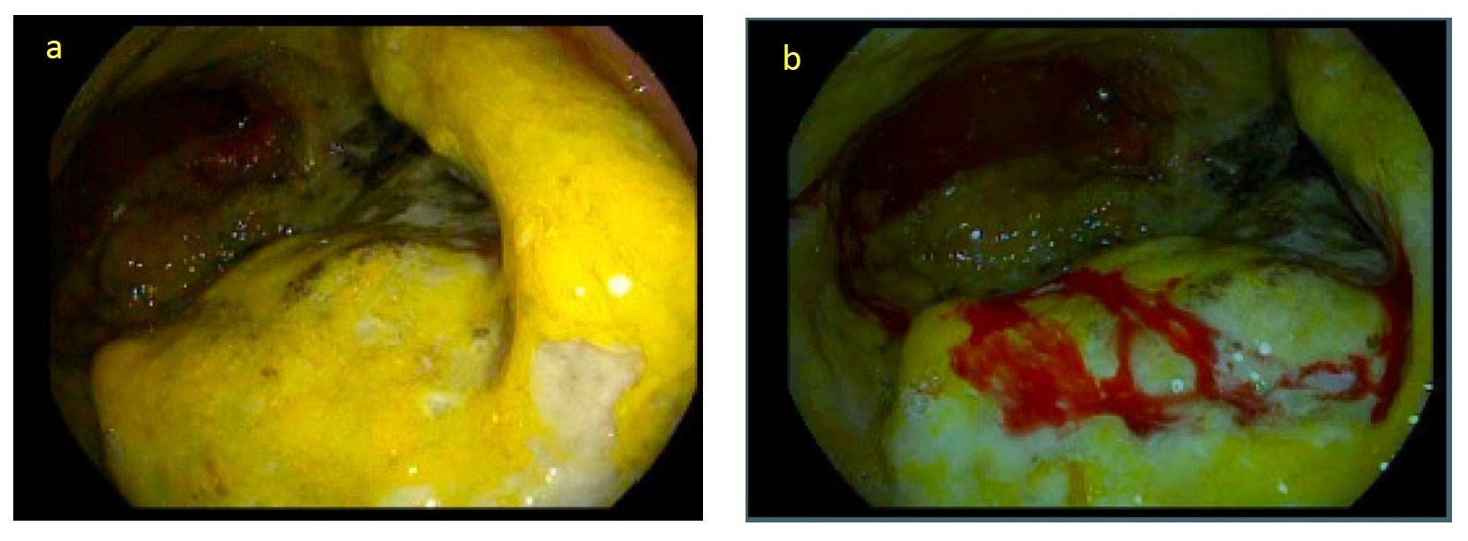
At this presentation, he reported a 15-pound weight loss and CT imaging demonstrated significant right colon wall thickening with a 19cm “mass-like” lesion. A subsequent colonoscopy showed a large, ulcerated, partially obstructing right colon mass consistent with malignancy. Histology demonstrated a high-grade B-cell lymphoma that was CD20 positive by immunohistochemical staining. Unfortunately, the patient was discharged from our facility at his request before definitive therapy could be undertaken.
Two weeks later, he presented to a different ED with bloody diarrhea, abdominal pain, and vomiting and was found to have perforation of the cecum with free air. He underwent an exploratory laparotomy with a stormy postoperative course and eventually died from post-surgical complications.
Discussion: Although a primary colonic lymphoma is exceedingly rare, especially in the young population, this case is instructive as it is common to overlook malignancy in the young that presents with gastrointestinal symptoms. The patient was seen in 2 separate hospitals and treated symptomatically even when he presented with profound anemia (hemoglobin of 4) and an abnormal CT scan of the right colon. Presentation of the disease can vary, however, should be considered and recognized in younger patients to avoid delays in proper management, which could lead to severe complications, as illustrated by this case. Given its rarity, no large trials have been conducted to evaluate optimal treatment.

Disclosures:
Hassan Zreik, MD, Parth M. Patel, MD, Sruthi Ramanan, MD, Harjinder Singh, MD, Shamik Parikh, MD, Merritt Bern, MD. A0120 - Primary Colonic B-Cell Lymphoma in a Young Patient, ACG 2022 Annual Scientific Meeting Abstracts. Charlotte, NC: American College of Gastroenterology.
Henry Ford Jackson, Jackson, MI
Introduction: The development of extranodal NHL can be a diagnostic challenge despite the fact that 30% of the cases are extranodal and involve the gastrointestinal tract. The incidence of a primary colonic lymphoma is rare, especially in young patients.
Case Description/Methods: A 21-year-old man initially presented to our emergency department (ED) with abdominal pain, weakness, rectal bleeding, and anemia. Two months prior to this admission, he presented to an ED in Colorado with rectal bleeding and abdominal pain. He was diagnosed with gastroenteritis and discharged with antibiotics. He was evaluated in Ohio for syncope and profound anemia (hemoglobin of 4.2) two weeks later. A CT scan of the abdomen and pelvis demonstrated right colon wall thickening. He was diagnosed with inflammatory bowel disease and discharged on prednisone.
At this presentation, he reported a 15-pound weight loss and CT imaging demonstrated significant right colon wall thickening with a 19cm “mass-like” lesion. A subsequent colonoscopy showed a large, ulcerated, partially obstructing right colon mass consistent with malignancy. Histology demonstrated a high-grade B-cell lymphoma that was CD20 positive by immunohistochemical staining. Unfortunately, the patient was discharged from our facility at his request before definitive therapy could be undertaken.
Two weeks later, he presented to a different ED with bloody diarrhea, abdominal pain, and vomiting and was found to have perforation of the cecum with free air. He underwent an exploratory laparotomy with a stormy postoperative course and eventually died from post-surgical complications.
Discussion: Although a primary colonic lymphoma is exceedingly rare, especially in the young population, this case is instructive as it is common to overlook malignancy in the young that presents with gastrointestinal symptoms. The patient was seen in 2 separate hospitals and treated symptomatically even when he presented with profound anemia (hemoglobin of 4) and an abnormal CT scan of the right colon. Presentation of the disease can vary, however, should be considered and recognized in younger patients to avoid delays in proper management, which could lead to severe complications, as illustrated by this case. Given its rarity, no large trials have been conducted to evaluate optimal treatment.

Figure: Figure 1: a and b. An ulcerated partially obstructing large mass in the ascending colon. The mass was circumferential, measured 10 cm in length and 10 cm in diameter. Oozing was present.
Disclosures:
Hassan Zreik indicated no relevant financial relationships.
Parth Patel indicated no relevant financial relationships.
Sruthi Ramanan indicated no relevant financial relationships.
Harjinder Singh indicated no relevant financial relationships.
Shamik Parikh indicated no relevant financial relationships.
Merritt Bern indicated no relevant financial relationships.
Hassan Zreik, MD, Parth M. Patel, MD, Sruthi Ramanan, MD, Harjinder Singh, MD, Shamik Parikh, MD, Merritt Bern, MD. A0120 - Primary Colonic B-Cell Lymphoma in a Young Patient, ACG 2022 Annual Scientific Meeting Abstracts. Charlotte, NC: American College of Gastroenterology.

