Back


Poster Session D - Tuesday Morning
Category: Liver
D0590 - Hepatocellular Carcinoma With Para-Celiac Lymph Node Metastasis Presenting as Unknown Primary Neoplasm in the Post-Liver Transplant Setting
Tuesday, October 25, 2022
10:00 AM – 12:00 PM ET
Location: Crown Ballroom

Has Audio
- MZ
Muhammad Adnan Zaman, MD
Conemaugh Memorial Medical Center
Johnstown, PA
Presenting Author(s)
Faisal Inayat, MBBS1, Rizwan Ishtiaq, MD2, Muhammad Adnan Zaman, MD3, Marjan Haider, MD4, Arslan Afzal, MD5, Haris Javed, MBBS1
1Allama Iqbal Medical College, Lahore, Punjab, Pakistan; 2St. Francis Hospital and Medical Center, Hartford, CT; 3Conemaugh Memorial Medical Center, Johnstown, PA; 4St. Joseph Mercy Hospital, Ypsilanti, MI; 5Woodhull Medical Center, Brooklyn, NY
Introduction: Metastatic HCC without primary liver lesion is an exceedingly rare clinicopathologic entity. Only a handful of case reports have been reported to date, mostly involving bony structures without primary liver cancer. To our knowledge, we hereby present the first case of metastatic HCC involving the para-celiac lymph nodes in the post-liver transplant setting.
Case Description/Methods: A 73-year-old male who previously underwent liver transplant for non-alcoholic steatohepatitis was admitted to GI for the evaluation of a suspicious pancreatic tail cyst. He denied acute GI symptoms. Laboratory studies revealed normal liver enzymes. CT abdomen and pelvis showed a 2-cm lesion located under the right hemidiaphragm, posterior to the right hepatic lobe. Subsequently, the patient underwent diagnostic EUS with biopsy for both lesions. EUS revealed an irregularly shaped oval cyst in the tail, round cysts in the body and neck, and a complex of small cysts in the pancreatic head. The 45 x 55-mm para-celiac
Discussion: To our research, this is the first reported case of metastatic HCC without primary liver lesion involving the para-celiac lymph nodes in a post-transplant patient. With regard to pathogenesis, one theory implicates the presence of ectopic liver tissue that transforms into HCC. The presence of micro-HCC that regresses spontaneously or gets destroyed by the immune system can also be a plausible explanation. Standard diagnostic protocol for HCC consists of imaging (CT or MRI) and/or biopsy. Even in patients with cirrhosis, the diagnostic modality of choice is imaging and liver biopsy is less frequently performed. Biopsy is recommended for atypical hepatic lesions on imaging or nonclassical enhancement patterns or detection in the absence of cirrhosis. No standardized treatment exists for metastatic HCC with unknown primary. This report illustrates that clinicians should consider HCC while evaluating patients for the primary origin of metastatic carcinoma.

Disclosures:
Faisal Inayat, MBBS1, Rizwan Ishtiaq, MD2, Muhammad Adnan Zaman, MD3, Marjan Haider, MD4, Arslan Afzal, MD5, Haris Javed, MBBS1. D0590 - Hepatocellular Carcinoma With Para-Celiac Lymph Node Metastasis Presenting as Unknown Primary Neoplasm in the Post-Liver Transplant Setting, ACG 2022 Annual Scientific Meeting Abstracts. Charlotte, NC: American College of Gastroenterology.
1Allama Iqbal Medical College, Lahore, Punjab, Pakistan; 2St. Francis Hospital and Medical Center, Hartford, CT; 3Conemaugh Memorial Medical Center, Johnstown, PA; 4St. Joseph Mercy Hospital, Ypsilanti, MI; 5Woodhull Medical Center, Brooklyn, NY
Introduction: Metastatic HCC without primary liver lesion is an exceedingly rare clinicopathologic entity. Only a handful of case reports have been reported to date, mostly involving bony structures without primary liver cancer. To our knowledge, we hereby present the first case of metastatic HCC involving the para-celiac lymph nodes in the post-liver transplant setting.
Case Description/Methods: A 73-year-old male who previously underwent liver transplant for non-alcoholic steatohepatitis was admitted to GI for the evaluation of a suspicious pancreatic tail cyst. He denied acute GI symptoms. Laboratory studies revealed normal liver enzymes. CT abdomen and pelvis showed a 2-cm lesion located under the right hemidiaphragm, posterior to the right hepatic lobe. Subsequently, the patient underwent diagnostic EUS with biopsy for both lesions. EUS revealed an irregularly shaped oval cyst in the tail, round cysts in the body and neck, and a complex of small cysts in the pancreatic head. The 45 x 55-mm para-celiac
Discussion: To our research, this is the first reported case of metastatic HCC without primary liver lesion involving the para-celiac lymph nodes in a post-transplant patient. With regard to pathogenesis, one theory implicates the presence of ectopic liver tissue that transforms into HCC. The presence of micro-HCC that regresses spontaneously or gets destroyed by the immune system can also be a plausible explanation. Standard diagnostic protocol for HCC consists of imaging (CT or MRI) and/or biopsy. Even in patients with cirrhosis, the diagnostic modality of choice is imaging and liver biopsy is less frequently performed. Biopsy is recommended for atypical hepatic lesions on imaging or nonclassical enhancement patterns or detection in the absence of cirrhosis. No standardized treatment exists for metastatic HCC with unknown primary. This report illustrates that clinicians should consider HCC while evaluating patients for the primary origin of metastatic carcinoma.

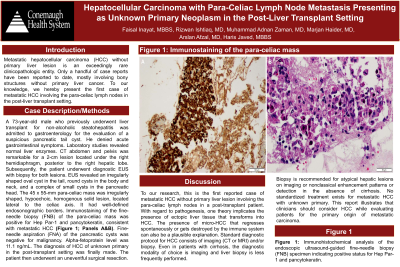
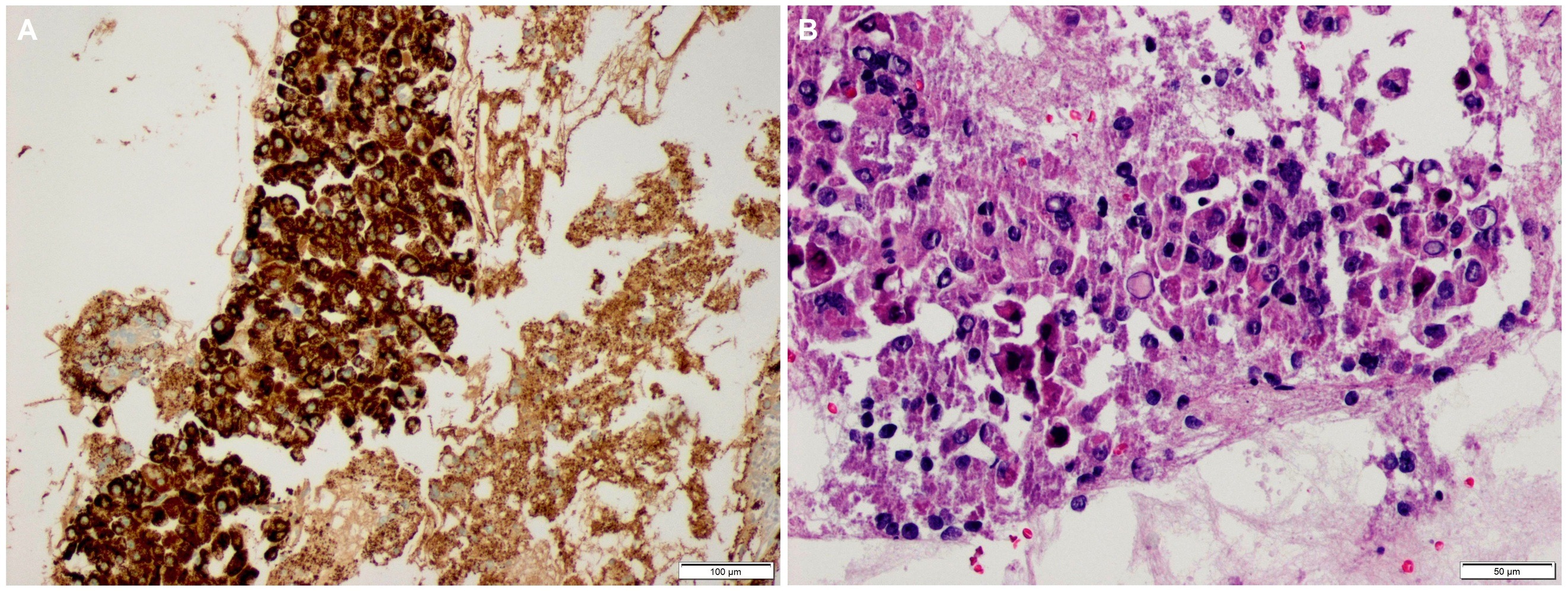
Figure: Figure 1. Immunostaining of the para-celiac mass. Endoscopic ultrasound-guided fine-needle biopsy (FNB) specimen indicating positive status for Hep Par-1 and pancytokeratin.
Disclosures:
Faisal Inayat indicated no relevant financial relationships.
Rizwan Ishtiaq indicated no relevant financial relationships.
Muhammad Adnan Zaman indicated no relevant financial relationships.
Marjan Haider indicated no relevant financial relationships.
Arslan Afzal indicated no relevant financial relationships.
Haris Javed indicated no relevant financial relationships.
Faisal Inayat, MBBS1, Rizwan Ishtiaq, MD2, Muhammad Adnan Zaman, MD3, Marjan Haider, MD4, Arslan Afzal, MD5, Haris Javed, MBBS1. D0590 - Hepatocellular Carcinoma With Para-Celiac Lymph Node Metastasis Presenting as Unknown Primary Neoplasm in the Post-Liver Transplant Setting, ACG 2022 Annual Scientific Meeting Abstracts. Charlotte, NC: American College of Gastroenterology.
