Back


Poster Session E - Tuesday Afternoon
Category: Colon
E0134 - Colonic Lymphangiomatosis
Tuesday, October 25, 2022
3:00 PM – 5:00 PM ET
Location: Crown Ballroom

Has Audio
- AM
Adil S. Mir, MD
Virginia Tech Carilion School of Medicine, VA
Presenting Author(s)
Adil S. Mir, MD, Diana Dougherty, MD, William Abel, MD, Douglas Grider, MD, David Lebel, MD
Virginia Tech Carilion School of Medicine, Roanoke, VA
Introduction: Colonic Lymphangiomatosis
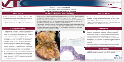
Case Description/Methods: A 73-year-old male with history of diabetes mellitus and chronic kidney disease underwent CT abdomen and pelvis with IV contrast (for the evaluation of hematuria and renal cyst), which showed incidental finding of free intraperitoneal air. The patient had a history of recent colonoscopy which showed a large concentration of ‘polyps’ in the sigmoid, not amenable for endoscopic removal. Subsequently the patient developed abdominal pain and nausea, and labs showed worsening leukocytosis 17K/uL. Repeat computed tomography (CT) abdomen revealed moderate free intraperitoneal air and innumerable colonic mucosal cysts of the very redundant sigmoid colon in the right upper quadrant. The patient underwent exploratory laparotomy, and although no perforation was seen, the sigmoid colon exhibited soft-tissue fullness, prompting partial sigmoid colectomy with primary anastomosis.
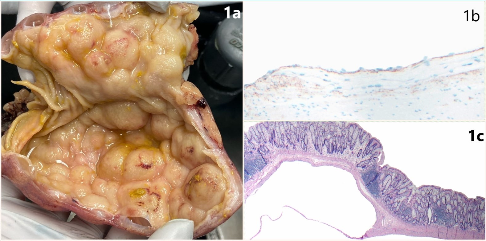
The resected specimen showed large contiguous well circumscribed and simple cystic structures (up to 3 cm size) giving a sessile “polypoid” appearance of the mucosa (Figure 1a). Histologic sections demonstrated overlying unremarkable colonic mucosal surface without epithelial dysplasia or serrated neoplasia, large cystic spaces extending into the submucosa, focally separated by thin fibrous septae (Figure 1b). The contiguous cystic spaces within the muscularis propria were noted to be lined by a bland and attenuated cell layer, showing immunoreactivity for CD31, CD34 and D2-40 (podoplanin – an immunostain defining lymphatic space endothelium) (Figure 1c); compatible with dilated lymphatic spaces and consistent with colonic lymphangiomatosis (CL).
Discussion: CL should be considered as a differential in patients with multiple aggregated mucosal lesions that appear as polyps on endoscopy. Speculation on etiology has ranged from developmental (failure to establish connections to lymphatic drainage sites), or seen secondarily after trauma, prior surgeries, radiation, lymphatic obstruction possibly secondary to adjacent mass lesion, or previous inflammatory processes. Free intraperitoneal air may suggest impending clinical decline in such patients. Surgical resection is the treatment of choice in symptomatic patients.
References:
Chung WC, Kim HK, Yoo JY, et al. Colonic lymphangiomatosis associated with anemia. World J Gastroenterol. 2008;14(37):5760-5762

Disclosures:
Adil S. Mir, MD, Diana Dougherty, MD, William Abel, MD, Douglas Grider, MD, David Lebel, MD. E0134 - Colonic Lymphangiomatosis, ACG 2022 Annual Scientific Meeting Abstracts. Charlotte, NC: American College of Gastroenterology.
Virginia Tech Carilion School of Medicine, Roanoke, VA
Introduction: Colonic Lymphangiomatosis
Case Description/Methods: A 73-year-old male with history of diabetes mellitus and chronic kidney disease underwent CT abdomen and pelvis with IV contrast (for the evaluation of hematuria and renal cyst), which showed incidental finding of free intraperitoneal air. The patient had a history of recent colonoscopy which showed a large concentration of ‘polyps’ in the sigmoid, not amenable for endoscopic removal. Subsequently the patient developed abdominal pain and nausea, and labs showed worsening leukocytosis 17K/uL. Repeat computed tomography (CT) abdomen revealed moderate free intraperitoneal air and innumerable colonic mucosal cysts of the very redundant sigmoid colon in the right upper quadrant. The patient underwent exploratory laparotomy, and although no perforation was seen, the sigmoid colon exhibited soft-tissue fullness, prompting partial sigmoid colectomy with primary anastomosis.
The resected specimen showed large contiguous well circumscribed and simple cystic structures (up to 3 cm size) giving a sessile “polypoid” appearance of the mucosa (Figure 1a). Histologic sections demonstrated overlying unremarkable colonic mucosal surface without epithelial dysplasia or serrated neoplasia, large cystic spaces extending into the submucosa, focally separated by thin fibrous septae (Figure 1b). The contiguous cystic spaces within the muscularis propria were noted to be lined by a bland and attenuated cell layer, showing immunoreactivity for CD31, CD34 and D2-40 (podoplanin – an immunostain defining lymphatic space endothelium) (Figure 1c); compatible with dilated lymphatic spaces and consistent with colonic lymphangiomatosis (CL).
Discussion: CL should be considered as a differential in patients with multiple aggregated mucosal lesions that appear as polyps on endoscopy. Speculation on etiology has ranged from developmental (failure to establish connections to lymphatic drainage sites), or seen secondarily after trauma, prior surgeries, radiation, lymphatic obstruction possibly secondary to adjacent mass lesion, or previous inflammatory processes. Free intraperitoneal air may suggest impending clinical decline in such patients. Surgical resection is the treatment of choice in symptomatic patients.
References:
Chung WC, Kim HK, Yoo JY, et al. Colonic lymphangiomatosis associated with anemia. World J Gastroenterol. 2008;14(37):5760-5762

Figure: Figure 1: (a) Gross image of the sigmoid colon, bottom aspect showing partial deroofing of the mucosa of a simple cystic cavity (b) Hematoxylin and Eosin (H&E) stain 40X (c) D2-40 positivity (brown membranous and cytoplasmic staining) 100X
Disclosures:
Adil Mir indicated no relevant financial relationships.
Diana Dougherty indicated no relevant financial relationships.
William Abel indicated no relevant financial relationships.
Douglas Grider indicated no relevant financial relationships.
David Lebel indicated no relevant financial relationships.
Adil S. Mir, MD, Diana Dougherty, MD, William Abel, MD, Douglas Grider, MD, David Lebel, MD. E0134 - Colonic Lymphangiomatosis, ACG 2022 Annual Scientific Meeting Abstracts. Charlotte, NC: American College of Gastroenterology.
