Back


Poster Session B - Monday Morning
Category: Biliary/Pancreas
B0077 - Metastatic Renal Cell Cancer with Pancreatic Mass
Monday, October 24, 2022
10:00 AM – 12:00 PM ET
Location: Crown Ballroom

Has Audio
- SC
Sushant Chaudhary, MD
St. Mary's Hospital
Middlebury, CT
Presenting Author(s)
Sushant Chaudhary, MD1, Subhash Chander, MD2, Sayeed Jaan Naqvi, MD2, Winston Magno, MD2, Praneet Wander, MD2
1St. Mary's Hospital, Middlebury, CT; 2St. Mary's Hospital, Waterbury, CT
Introduction: Pancreas is an uncommon site of metastatic disease. Almost 63% of pancreatic metastasis arise from renal cell cancer. We report a case of metastatic renal cell cancer to pancreas, with contralateral adrenal metastasis 15 years after a radical nephrectomy.
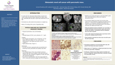
Case Description/Methods: 61 years old man with past medical history of renal cell cancer, status post left radical nephrectomy fifteen years ago, presented with dull pain in right lower abdominal for one day. He denied any other abdominal symptoms or weight loss. He smokes up to half a pack of cigarettes daily. His examination was unremarkable but he was noted to have elevated lipase (213U/L) and a bilirubin of 1.1 mg%. On computer tomography abdomen he had bulky, enhancing, soft tissue mass (~6.7 cm), with dilated pancreatic duct of 1.5 cm with right adrenal mass (Figure 1). On EUS, the lesion appeared well circumscribed and previously known pancreatic duct dilation was seen. Fine needle biopsy of the mass, which showed numerous atypical cells with round irregular nuclei with prominent nucleoli with clear cytoplasm suggestive of clear cell renal cell carcinoma (Figure 2). On immune -histochemistry, the tumor was positive for CD10, renal cell carcinoma antigen and PAX 8( Figure 2)
Discussion: Metachronous renal cell cancer to pancreas fifteen years after radial nephrectomy is rare. Our patient had pancreatic metastasis with adrenal involvement and presented with vague abdominal symptoms. .He was noted to have elevated lipase and dilation of main pancreatic duct. Due to a remote history of nephrectomy and the presenting complaints, pancreatic primary was suspected. However, on endoscopic ultrasound the patient was noted to have a well circumscribed lesion in the head of pancreas with dilated pancreatic duct(Figure 1). Regular borders and absence of pancreatic duct dilation suggested metastatic disease. Our patient had well circumscribed lesion but with a dilated duct.
Most pancreatic metastasis are reported to occur within ten years after treatment of the primary disease but our patient had metastasis fifteen years later without any loco-regional recurrence. Endoscopic ultrasound is extremely important and allows to evaluate the extent of the disease, involvement of portal vein/ superior mesenteric artery, presence of enlarged lymph nodes, which are a must for evaluating the surgical candidature besides providing tissue diagnosis which helped us establish the diagnosis of metastasis.

Disclosures:
Sushant Chaudhary, MD1, Subhash Chander, MD2, Sayeed Jaan Naqvi, MD2, Winston Magno, MD2, Praneet Wander, MD2. B0077 - Metastatic Renal Cell Cancer with Pancreatic Mass, ACG 2022 Annual Scientific Meeting Abstracts. Charlotte, NC: American College of Gastroenterology.
1St. Mary's Hospital, Middlebury, CT; 2St. Mary's Hospital, Waterbury, CT
Introduction: Pancreas is an uncommon site of metastatic disease. Almost 63% of pancreatic metastasis arise from renal cell cancer. We report a case of metastatic renal cell cancer to pancreas, with contralateral adrenal metastasis 15 years after a radical nephrectomy.
Case Description/Methods: 61 years old man with past medical history of renal cell cancer, status post left radical nephrectomy fifteen years ago, presented with dull pain in right lower abdominal for one day. He denied any other abdominal symptoms or weight loss. He smokes up to half a pack of cigarettes daily. His examination was unremarkable but he was noted to have elevated lipase (213U/L) and a bilirubin of 1.1 mg%. On computer tomography abdomen he had bulky, enhancing, soft tissue mass (~6.7 cm), with dilated pancreatic duct of 1.5 cm with right adrenal mass (Figure 1). On EUS, the lesion appeared well circumscribed and previously known pancreatic duct dilation was seen. Fine needle biopsy of the mass, which showed numerous atypical cells with round irregular nuclei with prominent nucleoli with clear cytoplasm suggestive of clear cell renal cell carcinoma (Figure 2). On immune -histochemistry, the tumor was positive for CD10, renal cell carcinoma antigen and PAX 8( Figure 2)
Discussion: Metachronous renal cell cancer to pancreas fifteen years after radial nephrectomy is rare. Our patient had pancreatic metastasis with adrenal involvement and presented with vague abdominal symptoms. .He was noted to have elevated lipase and dilation of main pancreatic duct. Due to a remote history of nephrectomy and the presenting complaints, pancreatic primary was suspected. However, on endoscopic ultrasound the patient was noted to have a well circumscribed lesion in the head of pancreas with dilated pancreatic duct(Figure 1). Regular borders and absence of pancreatic duct dilation suggested metastatic disease. Our patient had well circumscribed lesion but with a dilated duct.
Most pancreatic metastasis are reported to occur within ten years after treatment of the primary disease but our patient had metastasis fifteen years later without any loco-regional recurrence. Endoscopic ultrasound is extremely important and allows to evaluate the extent of the disease, involvement of portal vein/ superior mesenteric artery, presence of enlarged lymph nodes, which are a must for evaluating the surgical candidature besides providing tissue diagnosis which helped us establish the diagnosis of metastasis.

Figure: CT(A,B,C) and EUS(D,E) findings. Image A shows the mass in the Pancreatic head(arrow with red head) with a dilated pancreatic duct
seen in imageB( arrow with red head). Adrenal metastasis is seen in image C marked with a red arrow. On EUS, the mass appears well circumscribed( blue arrow, image D) with dilated pancreatic duct seen ( green arrow) in the head of pancreas in E.
seen in imageB( arrow with red head). Adrenal metastasis is seen in image C marked with a red arrow. On EUS, the mass appears well circumscribed( blue arrow, image D) with dilated pancreatic duct seen ( green arrow) in the head of pancreas in E.
Disclosures:
Sushant Chaudhary indicated no relevant financial relationships.
Subhash Chander indicated no relevant financial relationships.
Sayeed Jaan Naqvi indicated no relevant financial relationships.
Winston Magno indicated no relevant financial relationships.
Praneet Wander indicated no relevant financial relationships.
Sushant Chaudhary, MD1, Subhash Chander, MD2, Sayeed Jaan Naqvi, MD2, Winston Magno, MD2, Praneet Wander, MD2. B0077 - Metastatic Renal Cell Cancer with Pancreatic Mass, ACG 2022 Annual Scientific Meeting Abstracts. Charlotte, NC: American College of Gastroenterology.
